
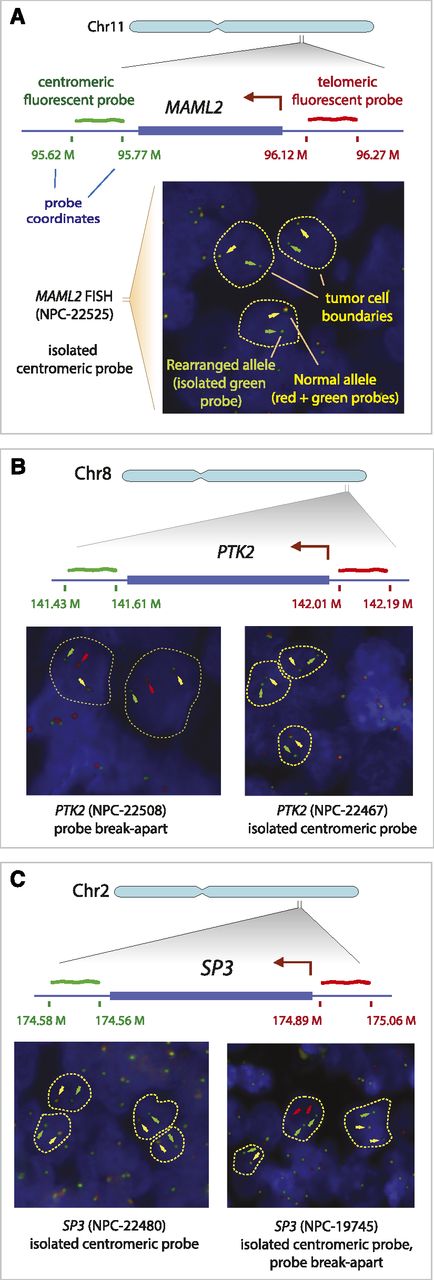
Recurrent rearrangements detected by fluorescent in situ hybridization of tissue microarrays. (A) Rearrangement of MAML2 in case NPC-22525. Probes were selected to flank centromeric (green probe) and telomeric (red probe) ends of the MAML2 gene. FISH images show cells with abnormal alleles. Approximate tumor cell boundaries are outlined with yellow dashed lines. Non-rearranged MAML2 alleles show colocalization of green and red probes (yellow arrows). Isolated green (or red) probes represent rearranged alleles and are marked by green (or red) arrows. (B) Two cases showing rearrangements of PTK2. (C) Two cases with a rearranged SP3 gene. NPC-22480 represents a core of a sequenced sample NPC-5421, which contains a rearranged SP3 allele, corresponding to the isolated centromeric probe (green).


