
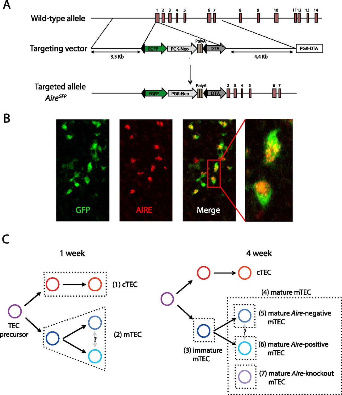
Generation of AireGFP/+ mice. (A) The genomic Aire locus (top), with the targeting construct (middle), and the targeted locus (bottom). Red rectangles with numbers indicate exons and black triangles indicate loxP sites. The PGK neo cassette in the targeting construct is followed by triple poly(A) signals to prevent further transcriptional elongation. (B) Immunofluorescence analysis of thymus sections of AireGFP/+ mice for GFP (green) and AIRE (red). (C) The basic scheme of TEC differentiation and the identification of individual TEC populations. Seven (1–7) distinct TEC populations were sorted from thymic tissue isolated from wild-type C57BL/6, AireGFP/+, and AireGFP/GFP mice (Supplemental Table 1; Supplemental Fig. 1). To ensure that the distinct mTEC subsets had fully differentiated during post-natal maturation in the presence of regular thymopoiesis and that these cells were sufficiently abundant for analysis (Irla et al. 2008), we collected the diverse mTEC populations from 4-wk-old animals, whereas cTEC and total mTEC were sorted from mice at 1 wk of age.


