
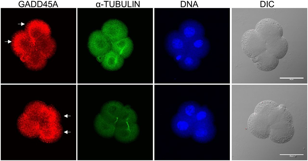
Figure 4.
GADD45A protein expression levels in two 4-cell mouse embryos. For each embryo (upper or lower panels), the confocal images were captured on the same z stack from an immunocytochemistry assay of GADD45A (Alexa-548, red) and alpha tubulin (Alexa-488, green). Arrows point to cells with greater expression of GADD45A. (DNA) DAPI fluorescence. (DIC) Differential interference contrast image. Scale bar, 50 μm.


