
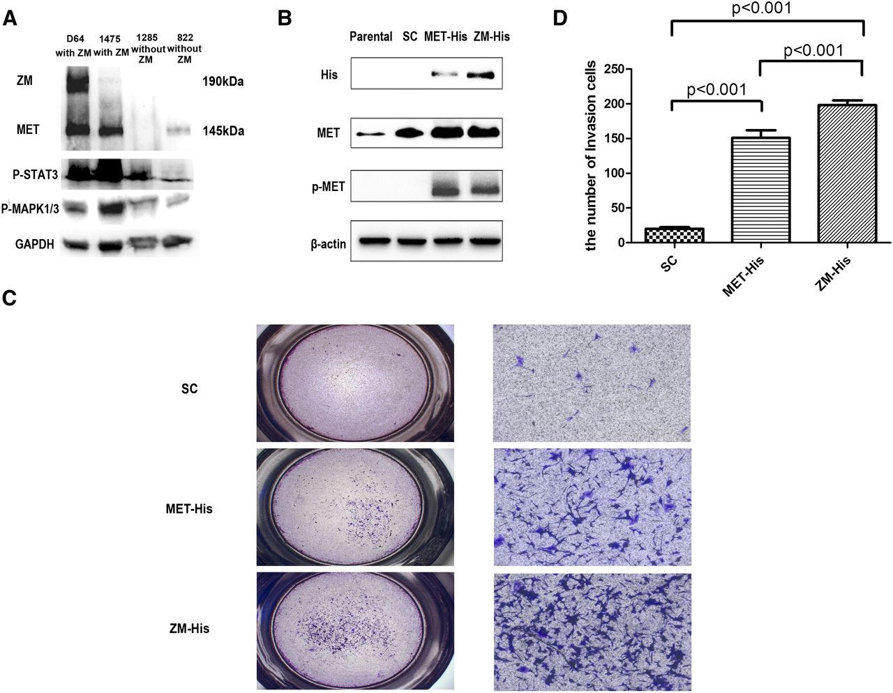
Immunoblot analysis and invasion assay used for oncogenic alteration detection of ZM fusion in vitro. (A) Immunoblot analysis of ZM-negative samples (822, 1285) and ZM-positive samples (1475, D64). Other than the band of wild-type (WT) MET at 145 kDa, D64 showed a distinct band at 190 kDa, in accordance with ZM fusion protein (ZM is 942 bp larger than WT MET at nucleotide level), which also hybridized with antibody to MET. STAT3 and the MAPK1/3 pathway were intensively activated in ZM-positive samples. (B) His tagged version of the CGGA_1475 ZM fusion was cloned into an adenovirus vector and stably expressed this protein in the U87MG GBM line. The MET endogenously expressed in U87MG is not phosphorylated at residue 1234/5. In contrast, exogenously expressed MET or ZM fusion harbors this phosphorylation. (C) Contrasted with scrambled, notably more U87 cells infected by ZM and MET adenovirus penetrated the Matrigel-coated transwell at 24 h after cells seeded. Meanwhile, the ZM group showed more an intensive invasion than did cells over expressing MET. (Left) 1×; (right) 10×, with scale bar, 200 μm. (D) The fold induction in migration relative to the SC (scramble control) group.


