
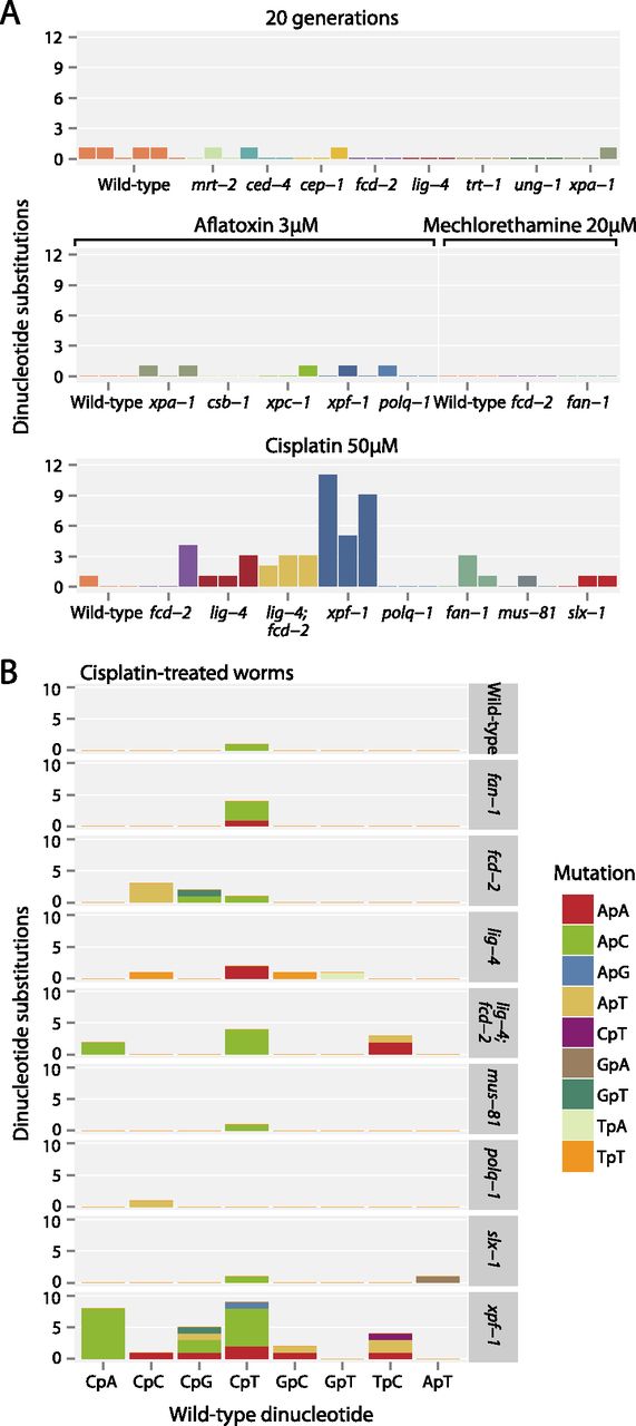
Figure 5.
Patterns of dinucleotide substitutions observed. (A) Numbers of dinucleotide substitutions in animals propagated for 20 generations and those exposed to the different carcinogens. (B) Dinucleotide substitutions in cisplatin-exposed animals. The sequences of the dinucleotides that are mutated are on the x-axis and what they are mutated to is denoted by the color of the bar.


