
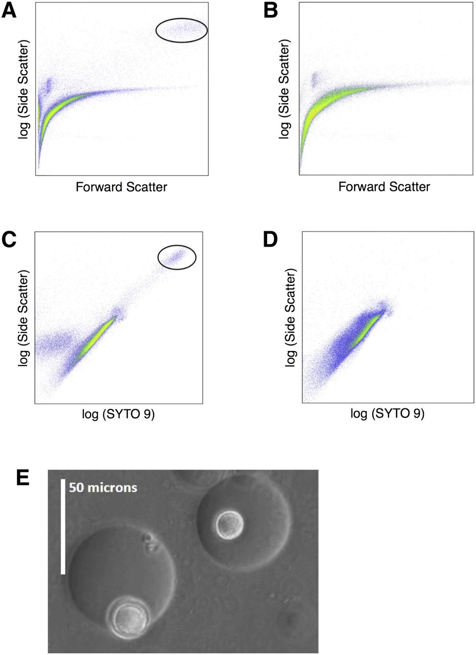
Figure 1.
Flow cytometry and light microscopy images of gel microdroplets (GMDs). (A,C) GMDs occupied with a colony are shown circled. (B,D) An unincubated sample that does not contain occupied GMDs. (E) Light microscopy image of GMDs containing a colony.


