
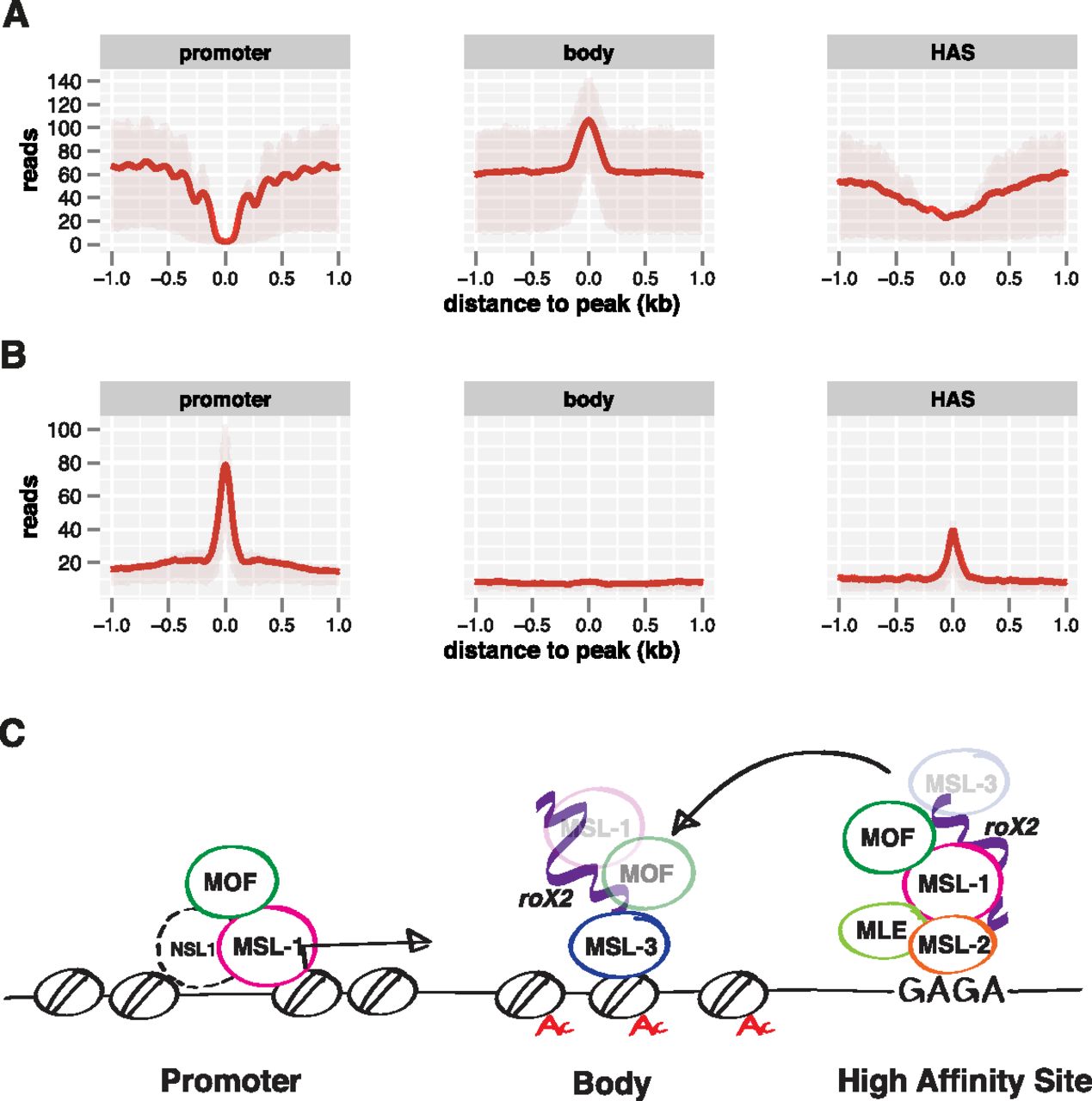
Figure 7.
Different modes of MSL protein binding in different chromatin contexts. (A) Nucleosome reads along MSL-1 peaks on promoters, MSL-3 peaks on gene bodies, and HAS as derived from MNase-seq data. Shaded areas above and below the solid lines describe the interquartile range of enrichment. (B) DNAse hypersensitivity along MSL-1 peaks on promoters, MSL-3 peaks on gene bodies, and HAS as derived from DNase-seq data (modEncode). Shaded areas on top and bottom of the solid lines describe the interquartile range of enrichment. (C) MSL complex architecture on three classes of binding sites as defined by high-resolution NGS mapping.


