
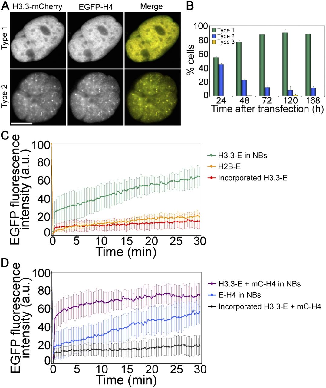
Expression of histone H4 accelerates the targeting of H3.3 to nuclear bodies and chromatin. (A) Colocalization of H3.3-mC and EGFP-H4 in NBs (type 2) and in chromatin (type 1). Scale bars, 10 μm. (B) Time course of detection of type 1, 2, and 3 H3.3-mC patterns after cotransfection with EGFP-H4 (mean ± SD of three experiments with more than 120 cells analyzed per experiment). (C) FRAP analysis of H3.3-EGFP (H3.3-E) in NBs or after incorporation into chromatin. The fluorescence recovery of chromatin-incorporated canonical core histone H2B-EGFP is shown as control (mean ± SD of seven to eight cells). (D) FRAP analysis of H3.3-EGFP in NBs or incorporated into chromatin in cells coexpressing mC-H4, and in cells expressing EGFP-H4 alone in NBs (mean ± SD of eight to 10 cells).


