
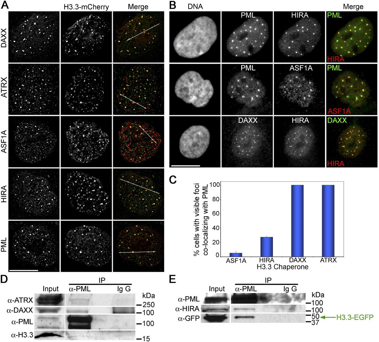
Epitope-tagged H3.3 and H3.3 chaperones are co-enriched at PML bodies. (A) Immunolocalization of DAXX, ATRX, HIRA, ASF1A, and PML in NBs, together with H3.3-mC (24 h after H3.3-mC transfection; deconvoluted images). In merged images, red is for H3.3-mCherry and green is for immunostainings. White lines in merged images delineate the zone of fluorescence signals quantified in Supplemental Figure 2A. (B) Immunolocalization of PML, HIRA, ASF1A, and DAXX at NBs in nontransfected cells. Scale bars, 15 μm. (C) Percentage of cells with NBs of ASF1A, HIRA, DAXX, and ATRX colocalizing with PML (mean ± SD of three countings in independent cell populations, with more than 300 cells per counting). (D) Coimmunoprecipitation of ATRX, DAXX, and endogenous H3.3 with PML, using anti-PML antibodies. (E) Coimmunoprecipitation of HIRA and H3.3-EGFP (detected with anti-GFP antibodies) with PML, using anti-PML antibodies, from cells overexpressing H3.3-EGFP (24 h after transfection).


