
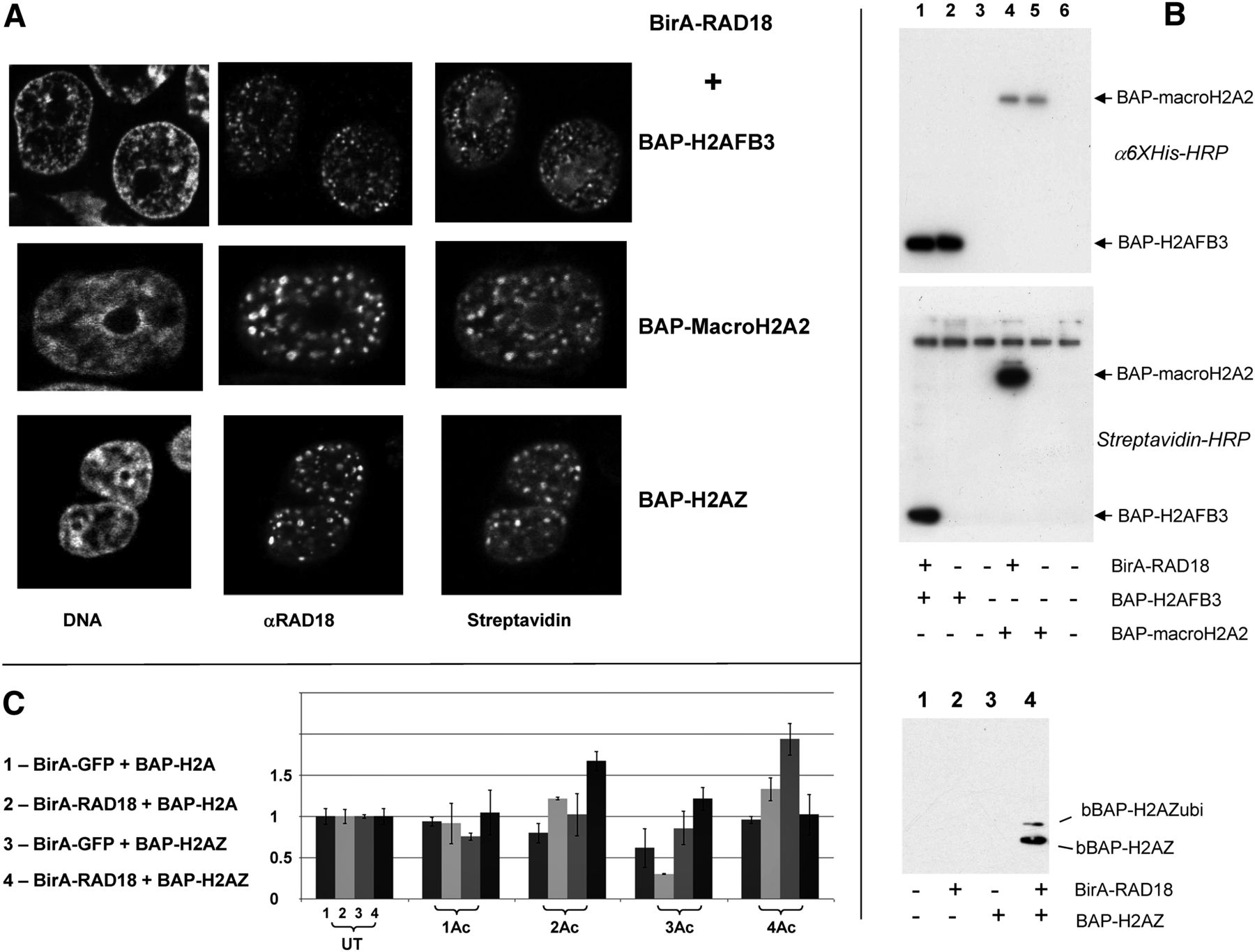
The pattern of H4 acetylation near RAD18 is different in the case of H2AZ-containing chromatin. (A) Alternative histones are biotinylated at the locations of RAD18. Confocal microscopy of 293T cells cotransfected by BirA-RAD18 and BAP-H2AFB3 (top), BAP-macroH2A2 (middle), and BAP-H2AZ (bottom). Shown are staining with Topro-3 (left), α−RAD18 (middle), and streptavidin (right). (B) Biotinylation signal is due to BAP-histone fusions. (Top and middle) Western blot analysis showing that a specific biotinylation signal appears only after BirA-RAD18 and BAP-macroH2A or BAP-H2AFB3 fusions are cotransfected. (Lanes 1,4) BAP-histone + BirA-RAD18; (lanes 2,5) BAP-histone; (lanes 3,6) control untransfected samples. (Top) α−6×His antibodies, (middle) streptavidin-HRP. (Bottom) Western blot analysis for the BAP-H2AZ fusion. The two forms of H2AZ (nonubiquitinated and ubiquitinated), both biotinylated in this experiment, are indicated by arrows. (C) Acetylation status of H4 N-terminal tails. The analysis was performed and is presented as described in the legend to Figure 2D. The heavy isotope-labeled cells were: (lane 1) BirA-GFP + BAP-H2A; (lane 2) BirA-RAD18 + BAP-H2A; (lane 3) BirA-GFP + BAP-H2AZ; (lane 4) BirA-RAD18 + BAP-H2AZ. The light isotope-labeled cells were always transfected with BirA-GFP + BAP-H2A.


