
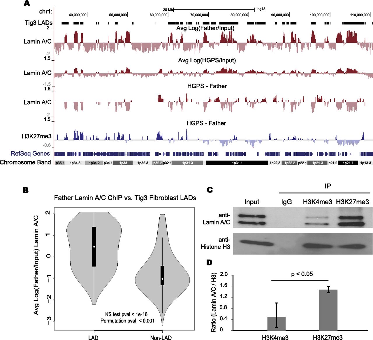
Dissociation of lamin A/C from heterochromatin regions in HGPS fibroblasts. (A) Regions of lamin A/C binding are shown as log (IP/Input) at 100-kb resolution for Father and HGPS fibroblasts along the left arm of chromosome 1. The previously classified LADs are shown above Father data (Tig3 LADs). Changes in lamin A/C binding and H3K27me3 association between Father and HGPS are shown below. (B) The distributions of lamin association signal in Father fibroblasts are shown for regions that were previously classified as LADs or non-LADs in Tig3 human lung fibroblasts. Lamin A/C ChIP-seq log ratios are significantly higher in LADs than in non-LADs as evaluated by a KS test and a circular permutation of the data (1000 permutations). (C) Western blotting analysis with anti-lamin A/C and anti-histone H3 antibodies in the immunoprecipitates with indicated antibodies. One percent of the Input and 10% of the immunoprecipitated proteins were run in each indicated lane. Three independent experiments were performed. One representative set is shown. (D) Quantification of the ratio of lamin A/C /Histone H3 of three independent replicates. There is a significant enrichment of lamin A/C in H3K27me3 ChIP compared with H3K4me3 ChIPs. P = 0.023.


