
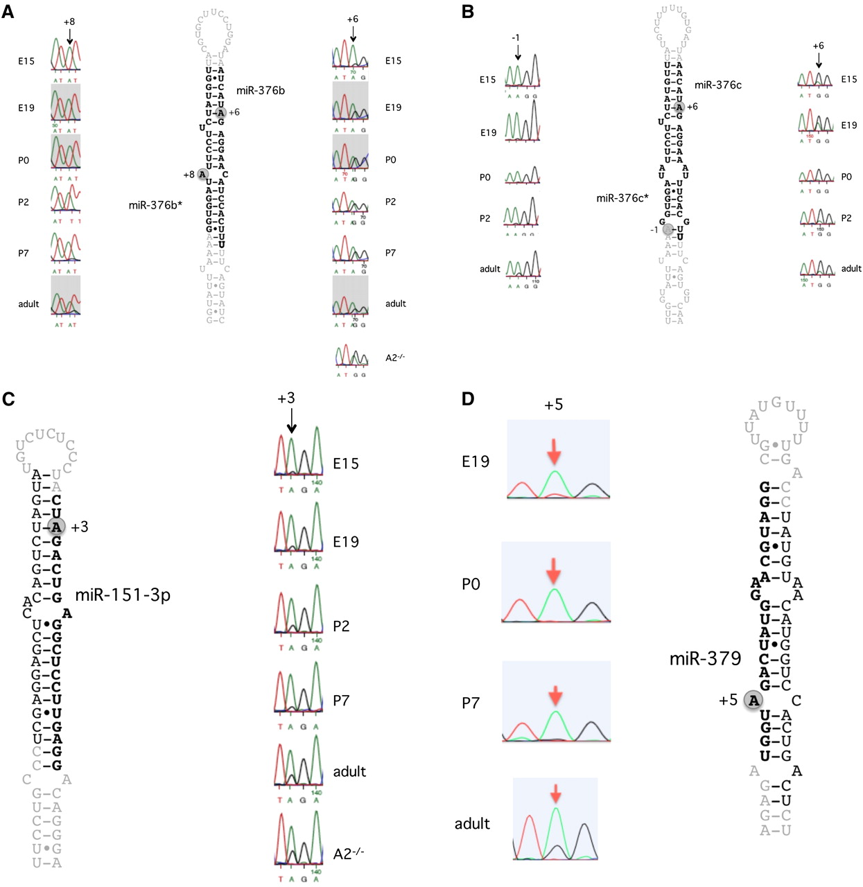
Figure 4.
Editing of pri-miRNAs at different developmental stages, detected by direct Sanger sequencing. RNA from whole mouse brain was amplified by RT-PCR followed by Sanger sequencing. Typical samples of biological triplicates are shown. The different developmental stages used for sequencing are indicated in each figure. The edited position is indicated by an arrow. (A) Editing of pri-miR-376b. (B) Editing of pri-miR-376c. (C) Editing of pri-miR-151-3p. (D) Editing of pri-miR-379-5p.


