
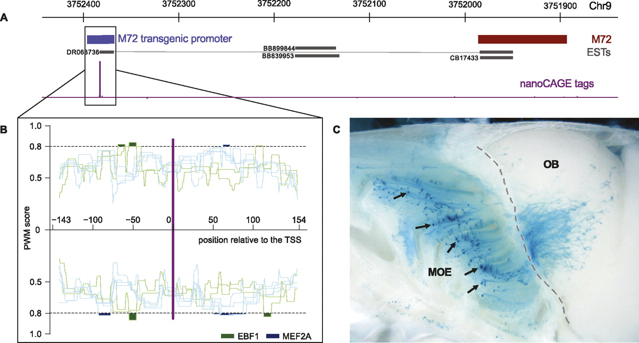
Transgenic reporter assay of the promoter activity of sequences flanking a nanoCAGE TSS. (A) Schematic depiction of the locus of Olfr160 (M72) on mouse chromosome 9. (Red) OLFR160 coding sequence; (blue) Olfr160 transgenic promoter; (purple) nanoCAGE tags; (black) ESTs. (B) Position weight matrix (PWM) scanning of the promoter sequences with matrices for EBF1 (green) and MEF2A (blue). Highlighted are the regions where the signal is above a 0.8 score threshold. (C) Whole mount of a mouse from transgenic line 7 shows X-gal-labeled cells predominantly in the middle domain of the MOE (black arrows). Labeled axons spread over a large domain in the middle aspect of the medial half-olfactory bulb, a probable consequence of the absence of an intact OR CDS in the transgene. The dotted gray line follows the shape of the cribiform plate separating the MOE from the olfactory bulb (OB).


