
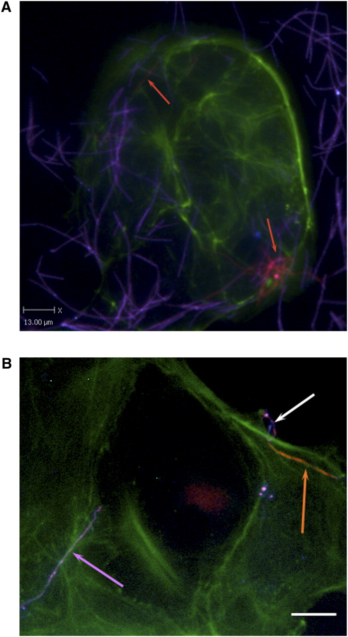
Representative differentially stained immunofluorescence image showing strain CC53 invading Caco-2 cells. (A) The differential staining method allows for delineation between bacteria that have penetrated the host cells (labeled for actin in green) to reside within them (orange, also indicated with orange arrows), and bacteria present on the outside of the cell (purple). CC53 shows a very long, fine, thread-like cell morphology. (B) Detail of CC53 invasion. (Top right) A representative CC53 cell in the process of invading the Caco-2 host cell (image is differentially stained as for 4A). The long, thread-like cells appear to penetrate host cells pole first. (Orange arrow) The CC53 cell that is internalized; (white arrow) the external portion of the same bacterial cell that has looped around on itself, demonstrating apparent flexibility. (Purple arrow) A single CC53 cell that has not invaded the host cells, for contrast. Bar, 15 μm. In the immunofluorescence micrograph, CC53 shows a very long, flexible cell morphology. (Green) Actin (Caco-2 cells); (orange) invasive and internalized bacteria; (purple) bacteria external to the cell.


