
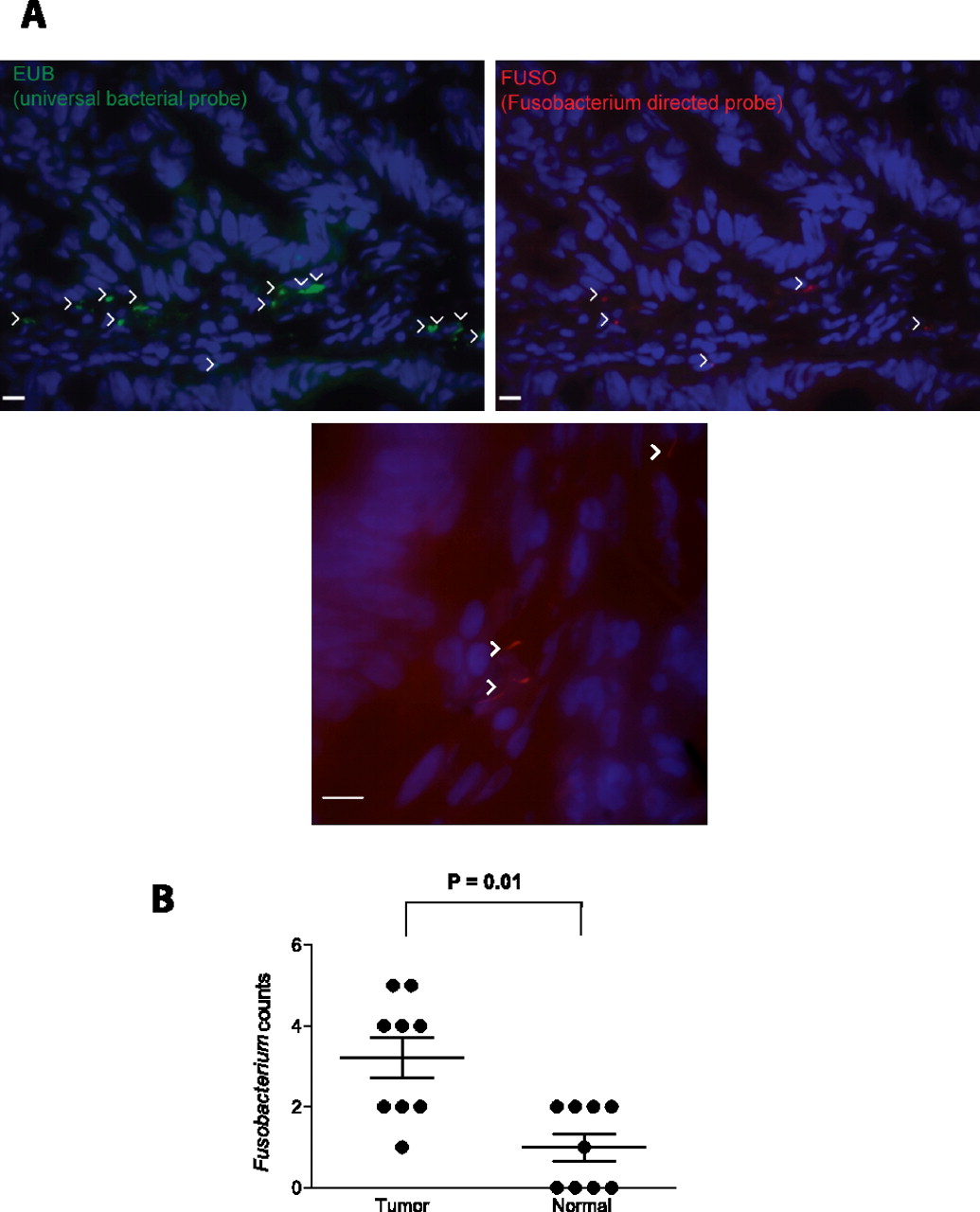
Fluorescence in situ hybridization (FISH) detects enrichment of Fusobacteria in colorectal tumors. (A) FISH using an Oregon-Green 488-conjugated ‘‘universal bacterial'' 16S rDNA-directed oligonucleotide probe (EUB338, green) (top left); and Cy3-conjugated Fusobacterium (FUSO, red) (top right and bottom center) 16S rDNA-direct oligonucleotide probe demonstrates the presence of bacteria and Fusobacterium within the colonic mucosa of colorectal tumor samples. Representative images are shown with a 10-μm scale bar in the lower corner of each panel; white arrowheads mark bacteria. Epithelial cell nuclei were stained with DAPI. (B) To determine whether Fusobacterium was enriched in tumor versus normal pairs, three random 40× fields were chosen for scoring by an observer blind to tumor/normal status, using selection criteria of mucosal tissue depth and a minimum of five bacteria visualized by the EUB338 probe per field. Each dot represents data from either a tumor or normal sample from nine tumor/normal paired cases. The mean, SEM, and P-values (calculated by a Wilcoxon matched-pairs signed rank test) are shown.


