
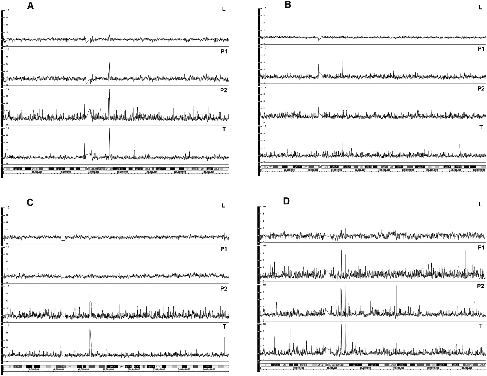
Figure 3.
High-level amplification in plasma and primary tumor DNA of breast cancer patients on follow-up. (A) Patient 44, amplification at 7q11.23 in tumor P1 and P2; (B) patient 27, amplification at 4q13.2 in tumor and P1; (C) patient 35, amplification at 5q13.2 in tumor and P2; and (D) patient 47, amplification at 10q11, showing two clear peaks (10q11.22 and 10q11.23) in tumor, P1 and P2. Top to bottom: L indicates normal leukocyte DNA; P1 and P2, paired plasma DNA samples; and T, FFPE tumor DNA.


