
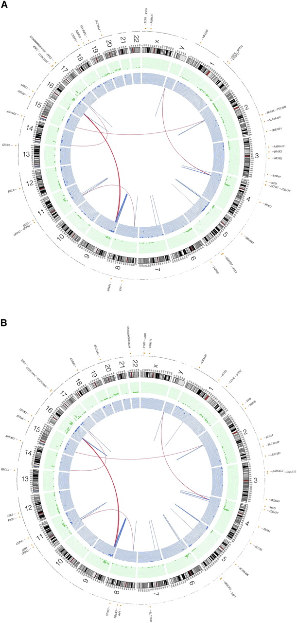
Figure 1.
Summary of lesions found in acral melanoma. Shown are primary and metastasis Circos plots (Krzywinski et al. 2009) of somatic mutations in the primary (A) and metastatic (B) tumors. The outer circle contains whole genome high-confidence SNVs (black dots) and nonsynonymous SNVs (orange dots; annotated with HGNC/Ensembl gene symbols). Copy number alterations are shown in the inner two plots (green circle shows gains and blue shows losses). Validated structural variations are depicted as links in the interior of the plot.


