
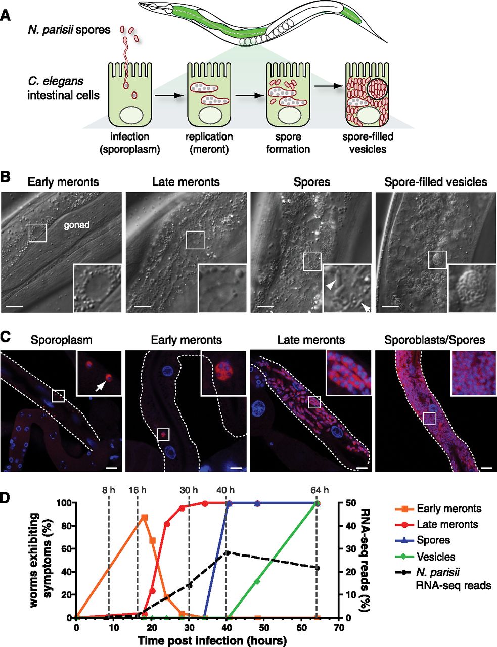
Characterization of N. parisii infection stages in C. elegans. (A) Diagram of infection stages. N. parisii infects C. elegans intestinal cells (green), where it grows intracellularly from small mononucleate sporoplasms to large, multinucleate meronts. Meronts develop into spores, which at late stages of infection can be enclosed within vesicles. (B) Representative DIC images of animals are shown displaying distinct infection-associated phenotypes with boxed areas enlarged and shown as insets. (Arrow) Small spore; (arrowhead) large spore. (C) N. parisii–specific FISH (red) and DAPI (blue) staining of animals at different stages of infection. Note: Although animals do not exhibit infection symptoms by DIC at 8 hpi, sporoplasm is visible by fluorescent in situ hybridization (FISH) at 8 hpi. (B,C) Scale bar, 10 μm. (D) The y-axis on the left indicates the fraction of animals in a population exhibiting specific symptoms of infection visualized by DIC (n > 22 animals assayed per time point) (see Supplemental Methods). RNA sample collection times are indicated on the graph: (1) 8 hpi, during the sporoplasm stage as observed by FISH; (2) 16 hpi, during the early meront stage; (3) 30 hpi, during the late meront stage; (4) 40 hpi, when spores have just started forming; and (5) 64 hpi, when Nematocida spores can be found within membrane-bound vesicles. The y-axis on the right indicates the contribution of N. parisii reads to total reads from RNA-seq analysis.


