
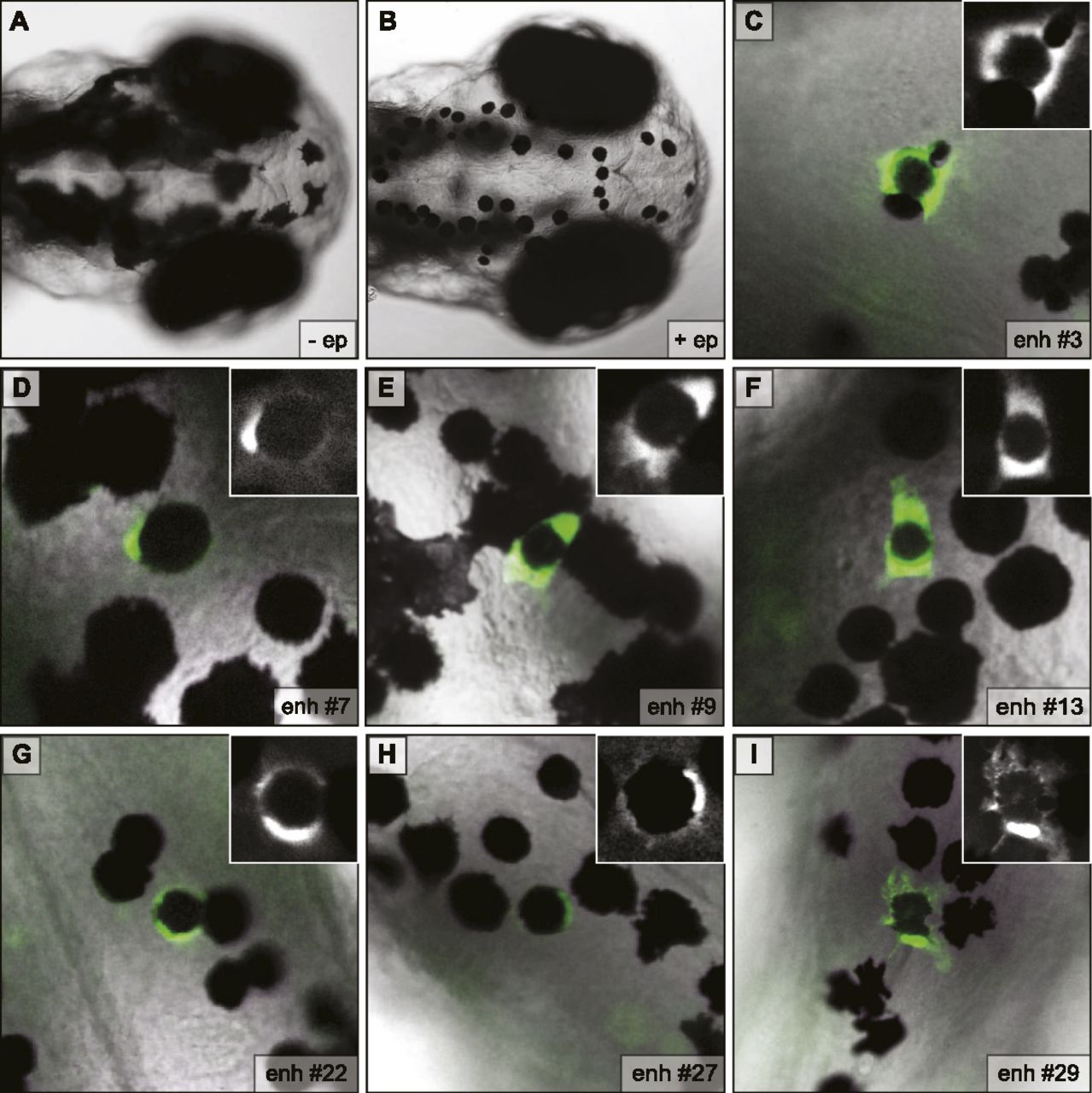
Figure 5.
Putative enhancers direct reporter expression in melanocytes of transgenic zebrafish embryos. (A) Dorsal view of melanocytes on the head of wild-type zebrafish embryo at 3 d post-fertilization (dpf). (B) Same view as A after treatment with epinephrine, which causes contraction of pigment granules to the center of the cell and enables the visualization of GFP at the periphery of melanocytes in transgenic embryos. (C–I) Representative images for all seven enhancers positive in this assay showing GFP-positive melanocytes in transgenic (mosaic) embryos at 3 dpf after treatment with epinephrine. Numbering is consistent with Figure 4.


