
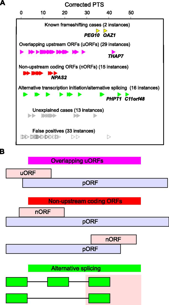
Figure 3.
Classification of dual coding regions. (A) Classification and PTS of 108 candidates. (B) Schematic organization of three major classes of dual coding. pORFs are shown as light blue bars and alternative frames as light pink bars. Splicing organization: green bars correspond to exons included in transcript variants, and lines indicate intronic regions excised during splicing.


