
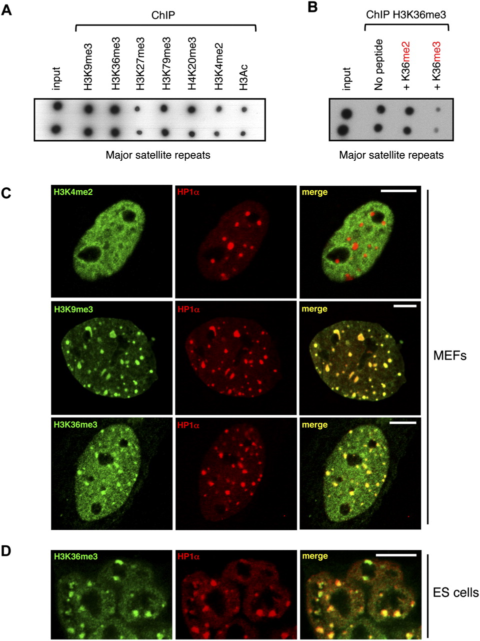
H3K36me3 is detected within pericentromeric heterochromatin regions. (A) Dot-blot analysis of DNA prepared from fetal brain chromatin immunoprecipitated with antibodies against the indicated histone modifications. (B) The specificity of H3K36me3 deposition within pericentromeric heterochromatin was tested by competition assay. Chromatin and antibodies against H3K36me3 were co-incubated with H3 peptides di- or trimethylated at lysine 36. Eluates were analyzed by dot blot. (C) Immunofluorescence confocal microscopy analysis of H3K36me3 distribution in mouse embryonic fibloblasts (MEFs), in comparison with other histone modifications. H3K4me2, H3K9me3, or H3K36me3 distribution is shown in green, whereas the pericentromeric heterochromatin marker HP1α is revealed as a red signal. Green and red signals were merged in the right panel (yellow indicates a colocalization). (D) Immunofluorescence analysis of H3K36me3 distribution in undifferentiated mouse ES cells. Scale bar, 10 μm.


