
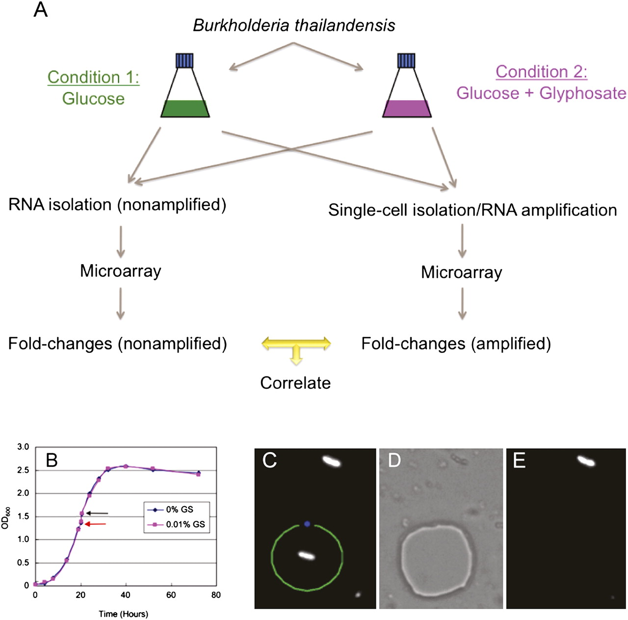
Single B. thailandensis cell isolation. (A) Experimental design for evaluating the single-cell transcript amplification method. B. thailandensis grown in two different conditions were used in large-scale (nonamplified) and single-cell level (amplified) microarray analysis. Fold-changes (between condition 1 and 2) of all genes detected from the nonamplified and amplified samples were then compared by correlation analysis. (B) Comparable growth curves of B. thailandensis in MG medium ± 0.01% GS (w/v) added at mid-log phase (red arrow) and harvested 30 min post-exposure (black arrow). (C) Fluorescent B. thailandensis cells were observed under 1000× magnification. The section of the membrane containing a single bacterium was drawn and cut by the focused laser (green line) and catapulted at a distance from the cell with unfocused low-intensity laser beam (blue spot), which aseptically catapulted and isolated the single cell into the lid of a 0.2-mL PCR tube containing the cell lysis buffer. (D) Bright-field mode showing the section of the membrane where the single bacterium had been. (E) Fluorescence mode confirming that the bacterium of interest has been transferred from the membrane slide to the PCR tube lid.


