
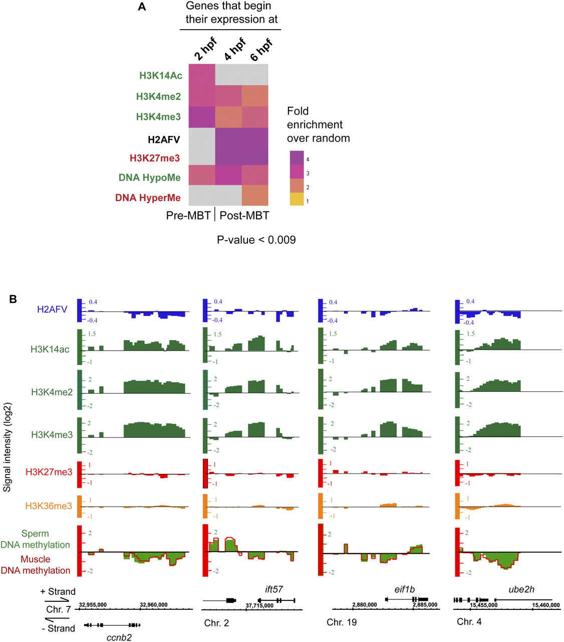
Certain chromatin modification patterns in sperm correlate with the timing of embryonic gene expression. (A) Heat map showing the correlations between genes that initiate expression at particular times during early development (2-hpf, 4-hpf, and 6-hpf columns from Mathavan et al. 2005) and particular histone modifications (or H2AFV enrichment) present at those genes in sperm (rows). Here, the color gradient shows the level of fold enrichment (over random) of a pairwise intersection between the list of expressed genes (Supplemental Table 18), and the list of genes with the respective histone modification/composition. A lack of significant intersection is depicted with a gray box. Genes expressed early/before MBT tend to have the highest levels of active histone modifications (acetylation and H3K4me2/3) in sperm, and those expressed after MBT tend to have repressive modifications (H3K27me3) in sperm. (B) Genes expressed before/early in MBT (3 hpf in zebrafish) are those that drive the cell cycle and promote metabolism, and are enriched with high levels of active histone modifications.


