
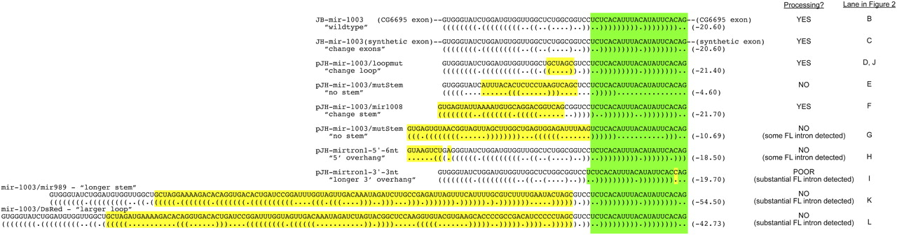
Figure 1.
Constructs used for structural analysis of mirtron biogenesis. Shown are sequence variants of the mir-1003 mirtron used for functional tests. (Green) The mature miRNA sequence; (yellow) the nucleotides differing from mir-1003. Their relative abilities to be processed in S2 cells are indicated (see also Fig. 2).


