
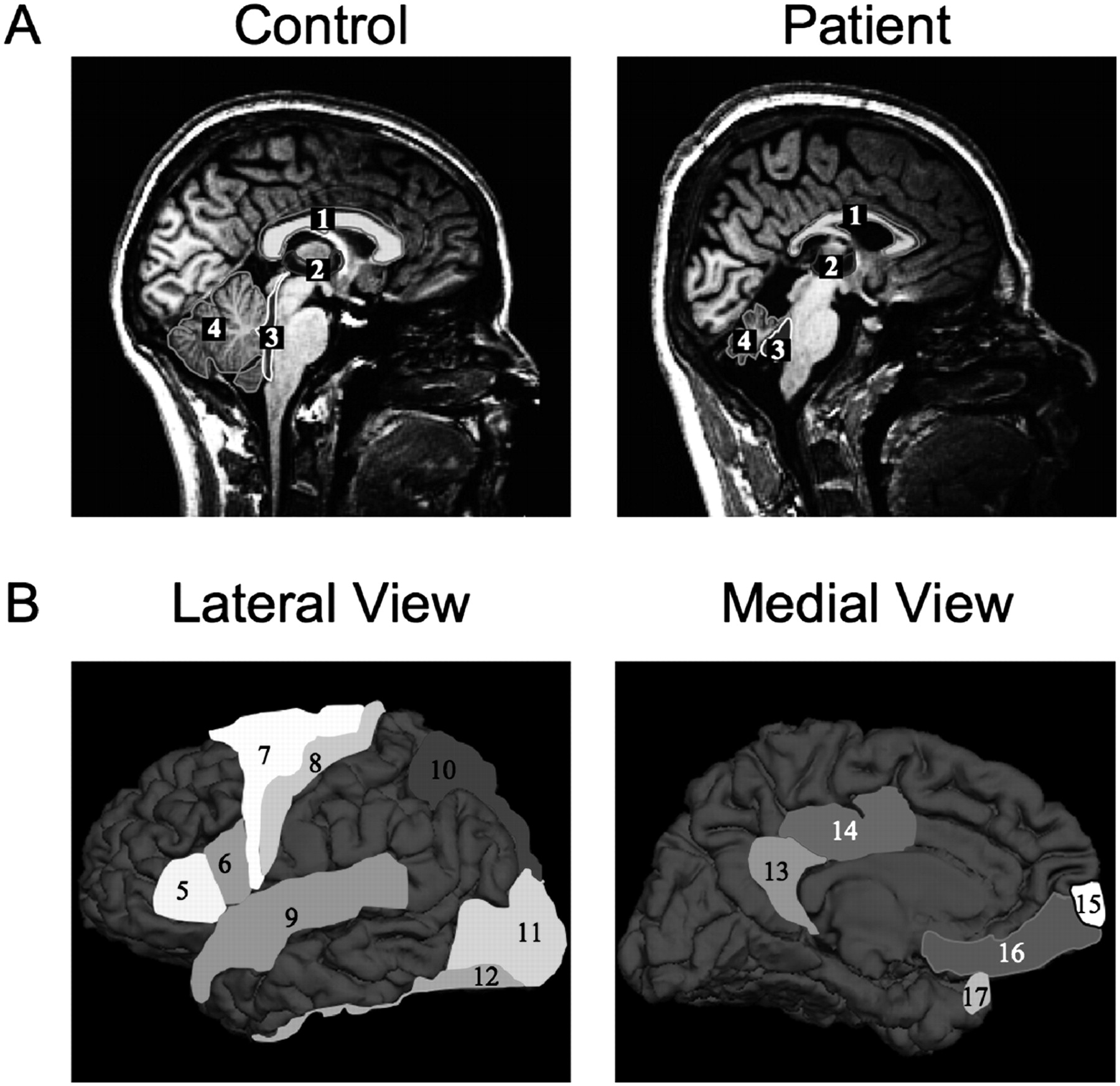
MRI-based morphological analysis of brain from affected and unaffected individuals. (A) Midsagittal MRI scans of a healthy control individual (left) and affected relative from Family B (right). The highlighted regions show areas where volumetric differences are readily visible: corpus callosum (1), third ventricle (2), fourth ventricle (3), and cerebellum (4). (B) Cortical regions with significant differences in morphometric parameters are displayed on a reference cortex, from lateral and medial view: BA45 (5), BA44 (6), BA6 (7), precentral (8), superior temporal (9), superior parietal (10), lateral occipital (11), fusiform (12), isthmus cingulated (13), posterior cingulated (14), frontal pole (15), medial orbitofrontal (16), and temporal pole (17). Additional details are provided in Supplemental Figure 2 and Supplemental Table 1.


