
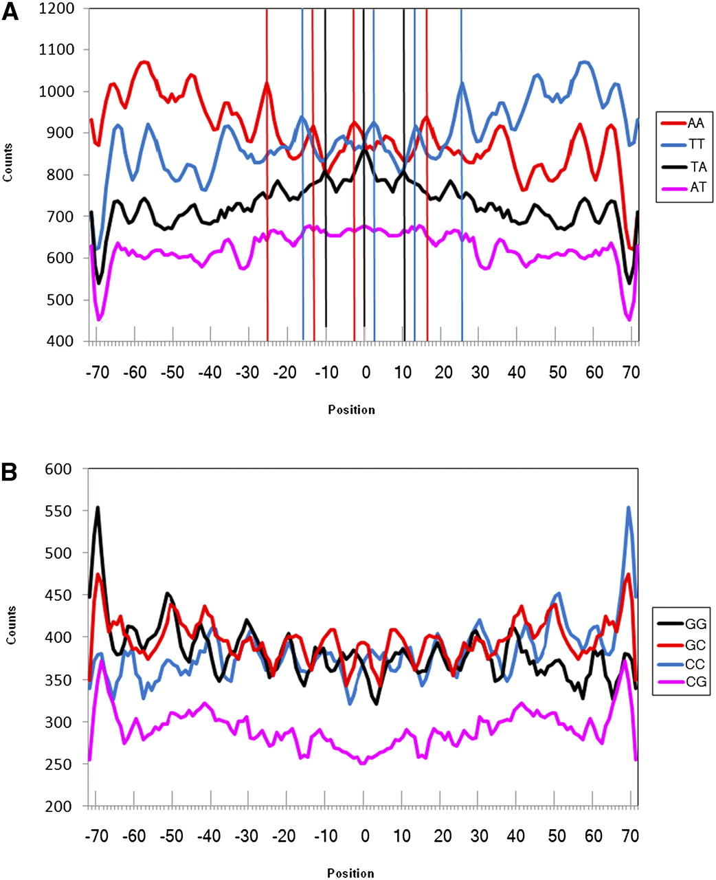
Figure 4.
Counts of individual dinucleotides along nucleosomal DNA for the studied nucleosomes from Albert et al. (2007). (A) Individual WW dinucleotides. (B) Individual SS dinucleotides. Vertical lines are drawn through the peaks for AA, TT, and AT dinucleotides that are clearly out of phase.


