
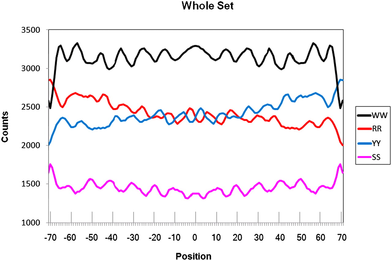
Figure 3.
Counts for combined dinucleotide distributions (smoothed by 3-points sliding average) for the well-phased H2A.Z nucleosomes used as a training set (according to Albert et al. 2007). Positions are for the aligned nucleosome DNA sequences. Position 0 coincides with the dyad symmetry of the nucleosome. Normalized frequency distributions can be found in Supplemental Figure 2.


