
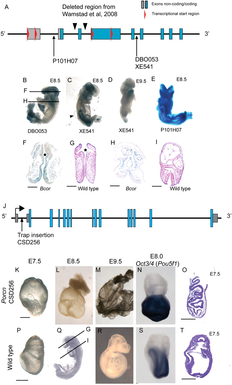
Analysis of Bcor and Porcn early embryo phenotypes. (A) Diagram of Bcor gene locus with locations of gene trap alleles, the conditional targeted allele as reported by Wamstad et al. (2008), and transcriptional start sites (red triangles). (B–E) mESC-derived embryos from three different Bcor GTs stained for lacZ expression. (B) Embryo from DB0053 cell line, used for histological analysis in F and H. Note the small head and heart, and truncated tail bud compared with controls (Q,R). (C) E8.5 mESC-derived embryo from XE541 cell line. Note the similar appearance and expression to DB0053 line. Also shown is a cyst (arrowhead) on the dorsal region of the embryo flanking the neural tube. (D) E9.5 Bcor mutant embryo stained for lacZ expression; of note is the severe posterior truncation, and small head and heart compared with wild-type controls (Q,R). (E) E8.5 embryo from P101H07 cell line exhibits exencephaly. (F) Transverse histological section of Bcor mutant embryo (B) head showing poor formation of optic pits (star), compared with an equivalent region from a wild-type stage-matched embryo (G). (H) Transverse histological section through heart field of Bcor mutant embryo (C) compared with equivalent region in stage-matched embryo (I). Bcor mutants (H) have a small symmetrical heart compared with the looped asymmetrical heart of controls (I). Also, Bcor mutants have an open neural tube compared with control embryos. (F–I) Scale bar, 100 μm. (J) Schematic of Porcn gene locus showing exons and GT insertion in first intron. (K–N) Porcn fully mESC-derived embryos. (P–S) Stage-matched wild-type embryos. (K) At E7.5, the Porcn null epiblast is highly folded and accumulates in the amniotic cavity compared to wild-type embryos (P). (L) At E8.5, Porcn mutants have no head, heart, allantois, or somite structures compared with stage-matched controls (Q). (M) At E9.5, Porcn mutant embryos consist of a yolk sac and small cyst-like structures, compared with wild-type controls that have a fully formed embryo body axis (R). (N) At E7.75 head-fold stage, Porcn mutant embryos fail to down-regulate Pou5f1 compared with stage-matched controls (S). (O) Hematoxylin and eosin-stained sagittal section along the presumed anterior posterior axis of Porcn mutants reveals details of highly folded epiblast and epiblast vesicles compared with the smooth epiblast of controls at E7.75 (T). Porcn mutants had no morphological node apparent in any of the sections from two mutant E7.5 embryos examined; also, the visceral endoderm does not appear to be displaced from the embryonic region of Porcn mutants. (K,P,O,T) Scale bar, 200 μm.


