
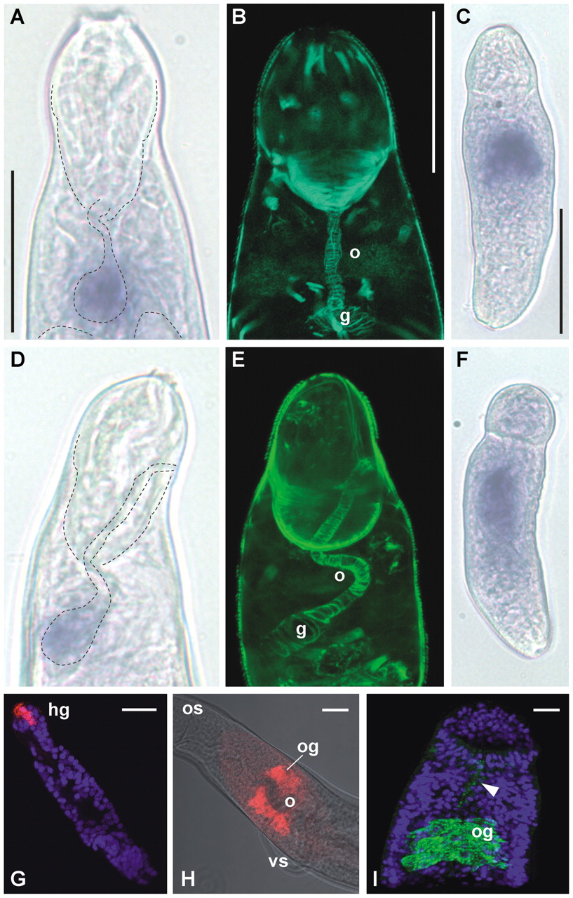
Whole-mount in situ hybridization (A,C,D,F–H) and immunocytochemistry (C,E,I) to identify the location of selected MEG gene expression products. (A) Focal expression of MEG-4.2 associated with the gut primordium of the nonfeeding infective cercariae, lying on its back. The fine dashed lines indicate the positions of the muscular head capsule and gut to aid interpretation. (B) The position of the embryonic esophagus and gut in a cercaria with similar orientation, revealed by Phalloidin staining of actin in the surrounding myocytes. (C) Increased expression of MEG-4.2 in the day 3 (skin stage) schistosomulum. (D) Focal expression of MEG-14 in a cercariae lying on its side, with structures outlined as in A. (E) A phalloidin stained day 3 schistosomulum in the same orientation to aid interpretation. Note that myocytes around the gut show only minimal expansion of the organ. (F) Increased expression of MEG-4.2 in the day 3 schistosomulum. (G) MEG-3.2 transcript expression sharply demarcated in the head gland (hg) of a day 10 (lung stage) schistosomulum. (H) Expression of MEG-4.1 in the esophageal gland (og) that envelops the ventral aspect of the esophagus (o). The gland lies adjacent to the ventral sucker (vs) distal to the mouth and oral sucker (os). (I) Confocal projection of MEG-4.1 protein detected in the male esophageal gland (og) using antibody to a synthetic peptide; traces of protein are also present in the esophageal lumen (arrowhead). The nuclei are counterstained with DAPI to reveal body outline. Magnifications: A,B,D,E all approximately the same, bar, 20 μm; C,F, bar, 50 μm; G–I, bars, 20 μm.


