
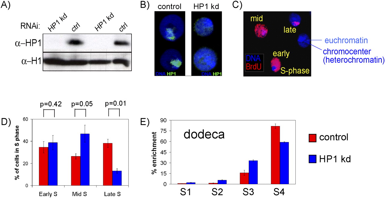
Depletion of HP1 by RNA interference. (A) Western blot detecting HP1 protein in untreated control cells (ctrl) and cells treated with dsRNA directed against HP1 (HP1 kd) for 7 d showing high efficiency of knockdown. H1 serves as a loading control for equal amounts of protein. (B) Cytological localization of HP1 by immunofluorescence with an antibody recognizing HP1 protein in Kc cells. HP1 localizes mainly to the chromocenter in control cells (left) but is detected only at very low levels in HP1 knockdown cells (right). (C) Cytological analysis of replication timing. Kc cell nuclei with three different patterns of BrdU incorporation after pulse-labeling are shown. (D) Quantification of the percentage of BrdU-positive nuclei with early, mid, and late S-phase pattern based on their BrdU staining in 486 control and 221 HP1-depleted BrdU-positive nuclei. Error bars, SEM between five biological repeats. P-values were calculated using the Wilcoxon rank sum test between five biological repeats. (E) Enrichments of BrdU containing DNA in four FACS sorted fractions (S1–S4) as quantified by real-time PCR. S1 represents the earliest and S4 the latest S-phase fraction as measured by DNA content. Here the dodeca pericentric repeat sequence is tested (see text). Error bars, SD between three biological repeats.


