
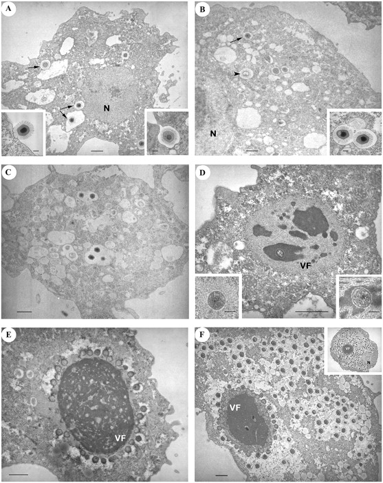
Progression of A. castellanii infection by Mimivirus over time. (A) T = −15 min: Some virus particles are inside vacuoles in the cytoplasm (arrows). (Left inset) Virus particles are sparsely found in contact with the cytoplasmic membrane. (Right inset) Phagocytosis of a virus particle. (B) T = 0: After 30 min of incubation with a large excess of virus (multiplicity of infection = 1000), the phagocytic vacuoles contain a mixture of empty (arrowheads) and intact (arrows) virus particles (probably not contributing to the measured viral transcripts). (Inset) Several particles can be gathered in the same vacuole. (C) T = 1.5 h. No major change is observed compared with T = 0. Both empty and intact viruses are still visible. (D) T = 3 h: The early virion factory appears as a gray structure, with a fibrous-like aspect, surrounding darker areas. A circular structure (the “seed”) is visible in one of these areas. (Left inset) In some cells, the “seed” is surrounded by the fibrous-like structure only. (Right inset) Higher magnification of the “seed” surrounded by dark matter. (E) T = 6 h: The fully mature virion factory now dominates the picture. Numerous particles are budding from its surface; most capsids are still empty. (F) T = 9 h: A large number of mature (hairy + DNA) virus particles are filling the cytoplasm. New particles are still produced by the virion factory. (Inset) T = 12 h: Ultimate stage of virion production. Panels: bar = 1 μm; insets: bar = 0.2 μm. (N) Nucleus; (VF) virion factory.


