
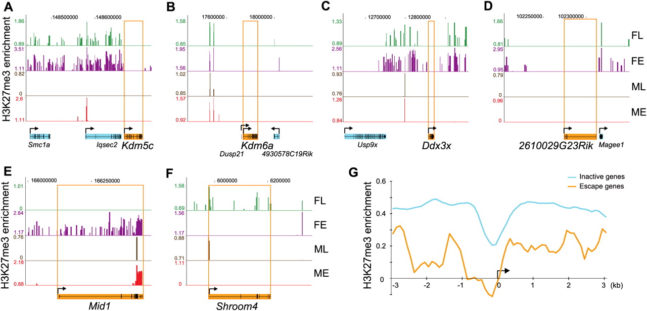
H3K27me3 is usually depleted at escape genes in female tissues. (A–D) Escape genes Kdm5c, Kdm6a, Ddx3x, and 2610029G23Rik are completely devoid of H3K27me3 in female tissues, while adjacent genes subject to X inactivation (e.g., Iqsec2, Magee1, Usp9x) are enriched. (E–F) Depletion in H3K27me3 at escape genes Mid1 and Shroom4 differs between tissues/developmental stages. (FL) Female liver; (FE) female embryos; (ML) male liver; (ME) male embryos. ChIP-chip peak files were uploaded in the UCSC Genome Browser (Mouse July 2007 [mm9] assembly). (Top) X chromosome coordinates. Escape genes are filled in orange, and inactive genes in light blue. (Arrow) Transcription direction. (G) H3K27me3 enrichment at the 5′ end of genes subject to X inactivation is higher than at escape genes in female liver. Average H3K27me3 enrichment for genes subject to X inactivation (366 genes; light blue curve) is compared to average enrichment for escape genes (10 genes; orange curve). Data are shown as log2 of the signal ratio between ChIP and input fractions for 3 kb upstream and 3 kb downstream of the transcription start site (black arrow).


