
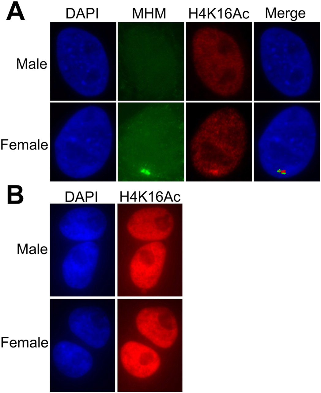
Figure 4.
(A) Colocalization in interphase chicken fibroblasts of the MHM RNA and histone 4 acetylated at lysine 16. (Left) DAPI-stained nuclei. (Second panel from left, green) RNA FISH showing the accumulation of MHM RNA near the MHM locus in females only. (Third panel from left, red) The accumulation of H4K16Ac at the same location. (B) The H4K16Ac staining of zebra finch cells does not have any specific pattern of signal accumulation. DNA was DAPI-counterstained. The signal strength of H4K16Ac staining in chicken (A) and zebra finch (B) is not comparable; the H4K16Ac signal in zebra finch was overexposed to illustrate the absence of even weak signal.


