
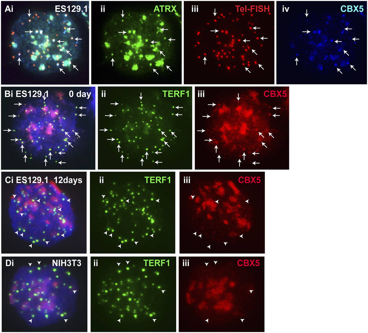
Colocalization of CBX5 with ATRX at the telomeres in ES cells. (A) A triple staining/immunofluorescence analysis showing colocalization of CBX5 (iv, blue) with ATRX (ii, green) at the pericentric heterochromatin (arrowheads) and telomeres (iii, red; indicated by telomere FISH). (B,C) CBX5 (Biii, red) colocalized with TERF1 (Bii, green) at the telomeres (examples shown by the arrows) in undifferentiated (0 day) interphase ES 129.1 cells. CBX5 signal remained apparent at the telomeres after 6 and 9 d of induction (data not shown). However, CBX5 localization (Ciii, red) at the telomeres (Cii, green) was greatly decreased 12 d following the induction of differentiation. (D) In somatic cells, e.g., NIH3T3, CBX5 (iii, red) localized mainly at pericentric heterochromation, but its association with the telomeres (arrowheads; ii, green) was below detection.


