
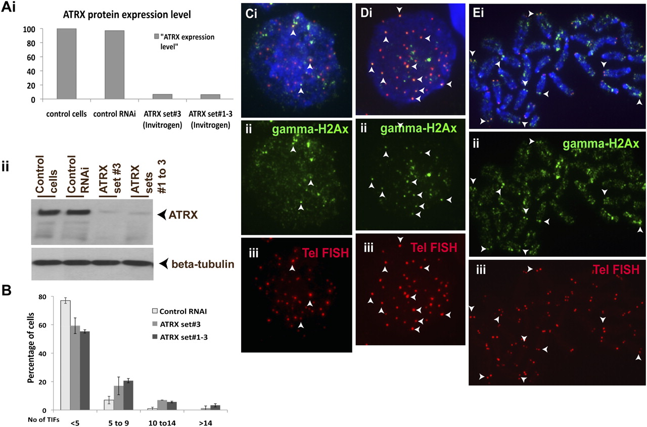
Effect of ATRX RNAi-knockdown on telomere integrity in mouse ES cells. (A) Western blot analysis of ES129.1 cells transfected with ATRX-specific RNAi oligonucleotide sets (set 3 and sets 1–3 from Invitrogen) using anti-ATRX and anti-beta-tubulin antisera. (i) Data are presented in histograms by normalization of the intensity of ATRX levels against the intensity of beta-tubulin levels. (ii) ATRX RNAi-depleted cells showed a significant reduction of ∼90% in ATRX protein level (arrows; lanes 3,4) 48 h after the transfection, compared with nontransfected cells (lane 1) and cells transfected with scrambled control RNAi oligonucleotides (lane 2). The equal loading of protein was achieved by normalization against the beta-tubulin level. (B) Induction of TIFs by ATRX-inhibition using ATRX RNAi oligonucleotide (set 3 and sets 1–3 from Invitrogen and set 2 from Ambion, respectively) for 48 h. Data are presented in histograms by subgrouping the cells according to the number of TIFs per cell (less than five 5 TIFs, five to nine TIFs, 10 to 14 TIFs, and more than 14 TIFs). A normal cell can contain one to two TIFs on average; thus, a threshold of four or more TIFs was used, as described in other studies (Hockemeyer et al. 2005). When transfected with ATRX-RNAi oligonucleotides, the number of cells with five or more TIFs (85 cells were counted for each experiment) by three- to fourfold (increased from 9.41% to as high as 29.8%–34.90%, with an average increase of 20.39%–25.49%). In this study, we only counted the number of TIFs in interphase ES129.1 cells although in some metaphase cells, TIFs were also present at the telomeres following RNAi-depletion of ATRX. We also performed RNAi-knockdown using ATRX-specific RNAi oligonucleotide sets purchased from Ambion, showing a significant reduction of ∼80% in ATRX protein level and a fivefold increase in the number of cells with five or more TIFs 48 h after the transfection (see Fig. S7). (C–E) Immunofluorescence analysis of ES129.1 cells subjected to 48-h knockdown with either control (C) or ATRX-specific (D,E) RNAi-duplex oligonuclotides using anti-gamma-H2AFX (Cii,Eii; green) antiserum. Increased number of TIFs was detected at telomeres (indicated by telomere FISH analysis; Diii,Eiii) in cells depleted of endogenous ATRX (arrowheads show some examples of TIFs).


