
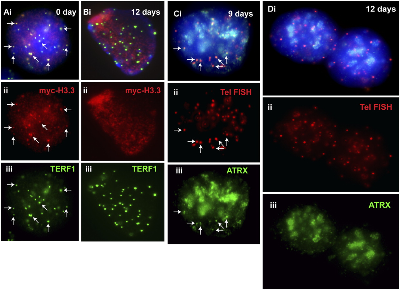
Cellular localization of MYC-H3.3 and ATRX in differentiated ES cells. (A,B) MYC-H3.3 construct was transfected into ES129.1 cells and induced with 1 μM doxycycline for 24 h MYC-H3.3 (Aii, red) colocalized with TERF1 (Aiii, green) at the telomeres (examples shown by the arrows) in undifferentiated (0 day) interphase ES 129.1 cells. MYC-H3.3 and ATRX signals remained apparent at the telomeres after 6 and 9 d of induction (data not shown). However, no enrichment of MYC-H3.3 (Bii, red) was detected at the telomeres (indicated by TERF1 staining; Biii, green) 12 d following the induction of differentiation. (C,D) Likewise, ATRX association at the telomeres was also not noticeably affected by the induction of differentiation for 6 and 9 d (Ciii, green), but ATRX (Diii, green) completely delocalized from the telomeres after 12 d of induction of cellular differentiation.


