
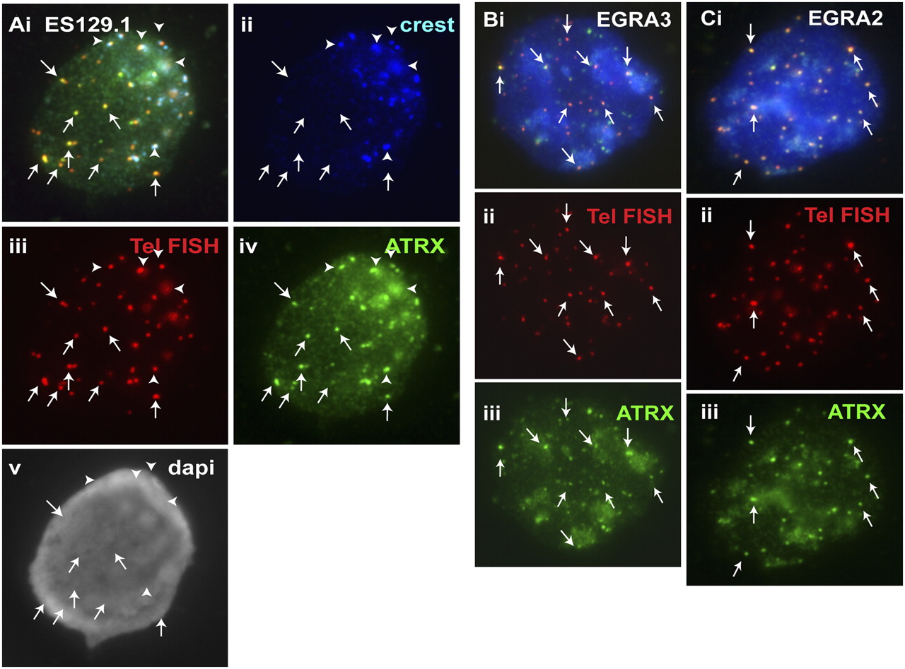
Figure 2.
Cellular distribution of ATRX in mouse ES cell lines ES129.1 and W9.5 and in mouse EG cell lines EGRA2 and EGRA3. (A) In interphase ES129.1 cells, a triple-staining immunofluorescence analysis showed clear colocalization of ATRX (iv, green) with telomere FISH signals (iii, red), without any CREST signal (arrows). ATRX localization at pericentric heterochromatin was indicated by its close proximity to the centromeric CREST staining (ii, blue; arrowheads). (B,C) In interphase EGRA3 and EGRA2 cells, ATRX (Biii,Ciii, green) was similarly enriched at the telomeres (arrows). Telomeric localization of ATRX (Biii,Ciii, green) was confirmed by telomere FISH (Bii,Cii, red).


