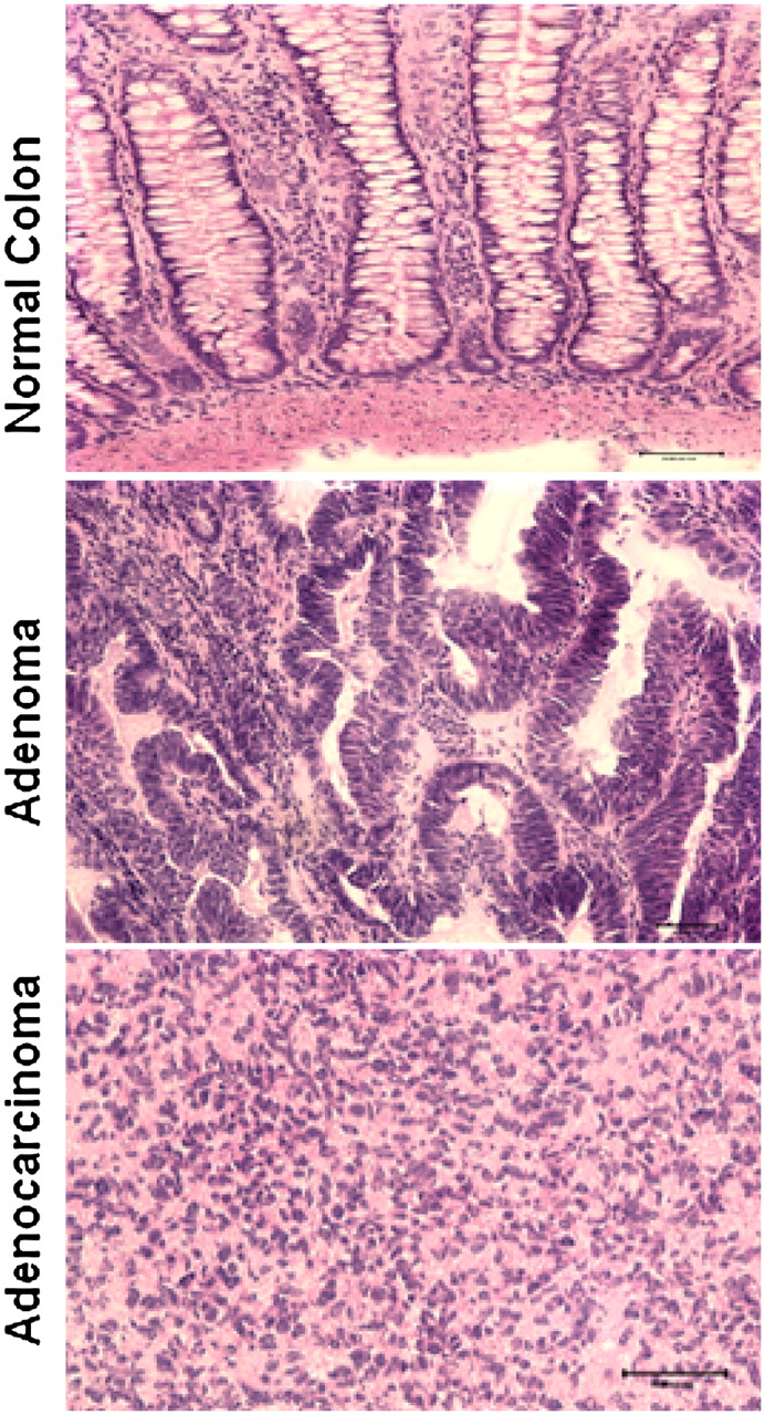
Copy number abnormalities in sporadic canine colorectal cancers

(Downloading may take up to 30 seconds. If the slide opens in your browser, select File -> Save As to save it.)

Click on image to view larger version.

Figure 2.
Figure 2.

Cryosectioning and H&E staining of dog colon tumor and normal tissue samples. The images represent a normal colon tissue (top), an adenoma (middle), and an adenocarcinoma (bottom).

This Article

  1. Genome Res. 20: 341-350

Preprint Server