
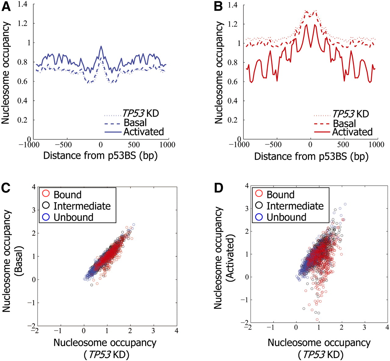
Nucleosomes are depleted from p53-bound sites upon activation of p53. (A) Nucleosome occupancy measurements per base pair, averaged across the p53-unbound sites (as defined in Fig. 1A), and shown along the 2000-bp region centered on each site. Results are shown for nonstressed MCF7 cells (Basal), MCF7 cells in which p53 was activated by Neocarzinostatin (Activated), and MCF7 cells with shRNA-mediated stable TP53 knockdown (TP53KD). Nucleosome measurements are shown as log-ratio between nucleosome sample and sonicated genomic DNA. (B) Same as in A, for the p53-bound sites. (C) Shown are nucleosome occupancy measurements (log-ratio between nucleosomal sample and sonicated genomic DNA) across the unbound (blue), intermediate-binding (black), and bound (red) p53 sites as defined in Figure 1A, averaged across all of the array probes that tile the 2000-bp region centered around the site. Results are shown for nonstressed MCF7 cells expressing endogenous wild-type p53 (Basal, y-axis) as well as cells with shRNA-mediated stable TP53 knockdown (TP53KD, x-axis). (D) Same as C, except that the y-axis corresponds to nucleosome measurements in MCF7 cells in which p53 was activated by Neocarzinostatin (Activated). Note the marked reduction of average nucleosome occupancy at many p53-bound sites (red group).


