
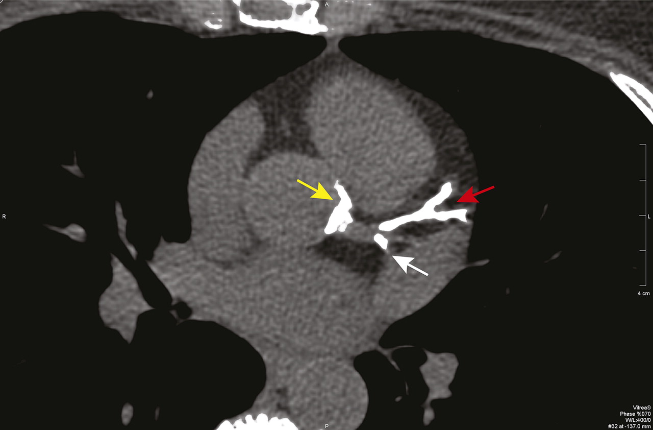
Figure 6.
Single axial slice of the coronary calcium scan from the patient described in Clinical Case 1 (Box 1) that shows severe calcification of the left anterior descending coronary artery (red arrow), the portion of the circumflex coronary artery within the imaging plane (white arrow), and the aortic root around the origin of the left main coronary artery (yellow arrow).


