
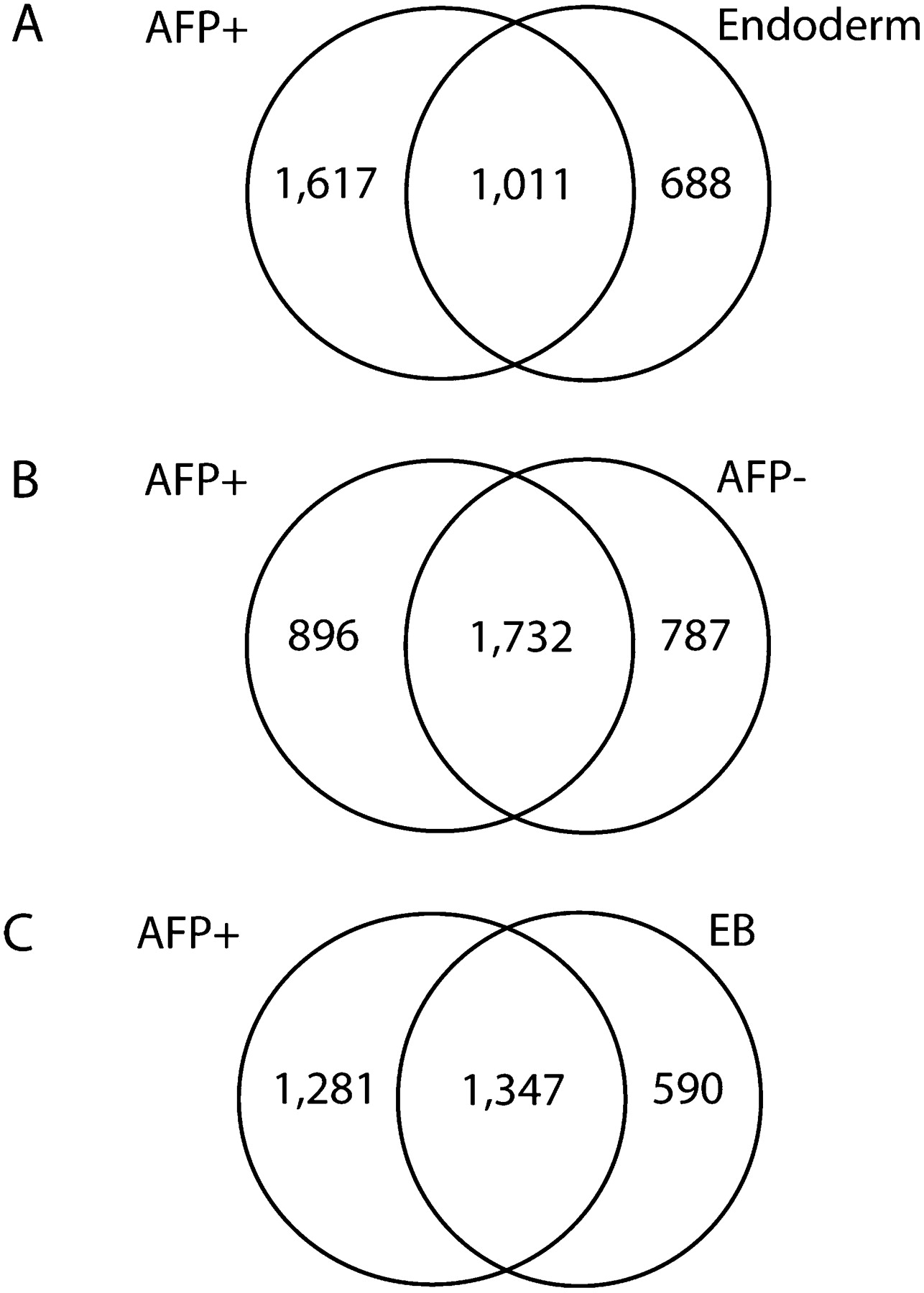
Figure 4.
DNA methylation changes occurring between naïve H9 hESCs and various differentiated samples. (A) Venn diagram for methylation differences between H9 hESC (5) and H9 AFP+ hESC-derived cells (7) and differences between H9 hESC (5) and H9 Endoderm (6). (B) Venn diagram for methylation differences between H9 hESC (5) and H9 AFP+ hESC-derived cells (7) and differences between H9 hESC (5) and AFP-negative hESC-derived cells (8). (C) Venn diagram for methylation differences between H9 hESC (5) and H9 AFP+ hESC-derived cells (7) and differences between H9 hESC (5) and H9 embryoid bodies (EB) (9). Library numbers are in parentheses.


