
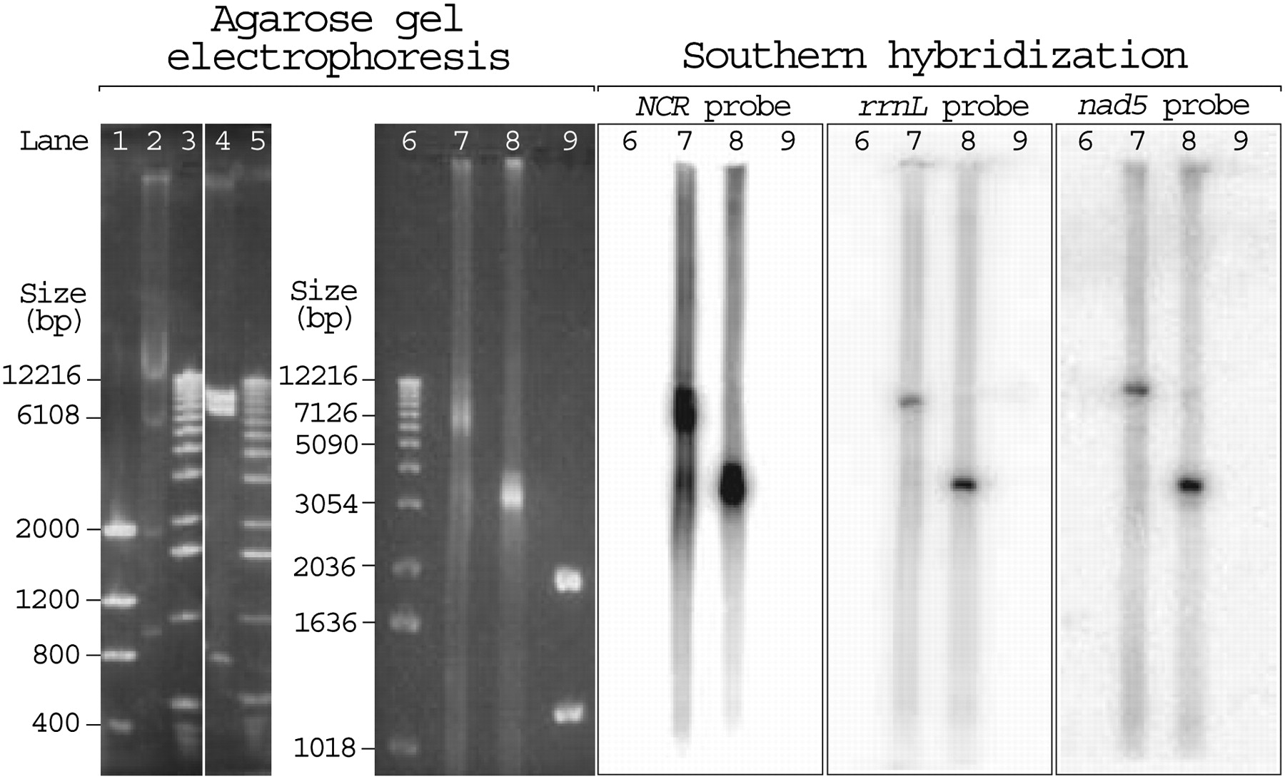
Agarose gel electrophoresis (left) and Southern hybridization (right) of the mitochondrial (mt) DNA of a fruitfly, D. melanogaster; a mouse, Mus musculus; and the human body louse, P. humanus. (Lanes 1,9) Two microliters low mass ladder DNA marker (Invitrogen); (lane 2) EcoRI-digested mtDNA extracted from182 mg of fruitflies; (lanes 3,5,6) 2 μL DNA molecular weight marker X (Roche); (lane 4) BamHI-digested mtDNA extracted from 36 mg of mouse liver tissues; (lane 7) undigested mtDNA extracted from 212 mg of human body lice; (lane 8) ApaI-digested mtDNA extracted from 212 mg of human body lice. The fruitfly and the mouse were experimental controls. As expected, the 19,517-bp mt chromosome of the fruitfly was cut into four fragments by EcoRI: ∼12,000, ∼5500, ∼1700, and ∼900 bp (lane 2); and the 16,300-bp mt chromosome of the mouse was cut into three fragments by BamHI: ∼8600, ∼7000, and ∼700 bp (lane 4). The undigested mtDNA of P. humanus migrated as a smear with a predominant size of 6000–9000 bp (lane 7), whereas the ApaI-digested mtDNA of P. humanus appeared as a block that ranges from 3000 to 4000 bp (lane 8).


