
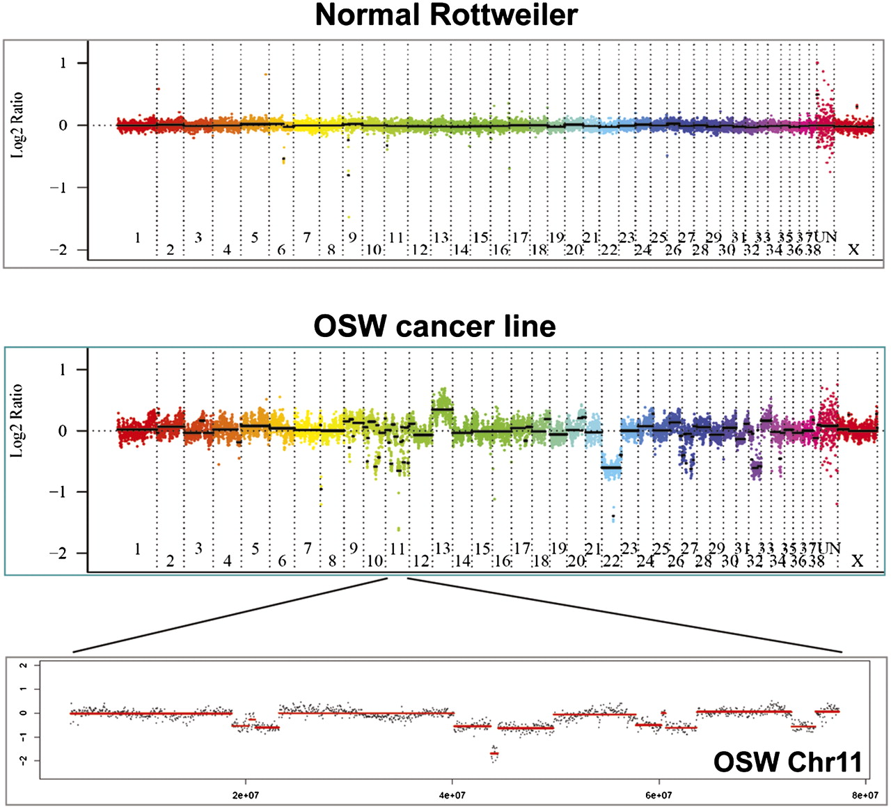
Whole-genome DNA copy number quantification of the OSW dog lymphoma cell line by aCGH. DNA was cohybridized with a reference pedigreed Boxer (in two colors) to an oligo array with <5 kb mean spacing. Copy number is shown as log2 ratios, with gains above the midline and losses below. The top panel shows the linearized whole-genome copy number of a normal Rottweiler and the middle panel that of OSW. (There is no Y chromosome data, and the last segment before the X is the unmapped genome assembly.) Note the complete or nearly complete gain of chr 13 and losses of chr 22 and chr 32. The bottom panel shows all of OSW chr 11, with seven large single-copy losses and one small two-copy loss. The P16 tumor suppressor gene lies within the two-copy loss region.


