
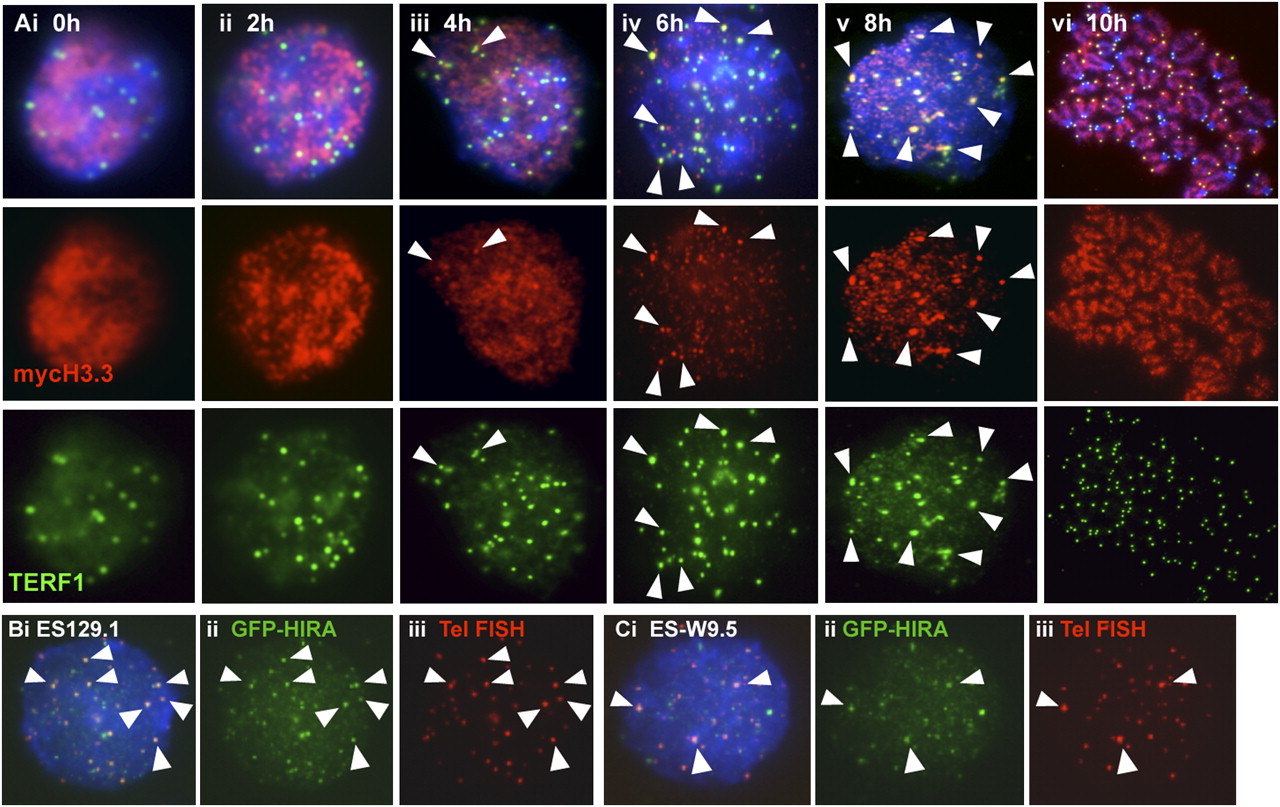
Immunofluorescence analysis of MYC-H3.3 telomeric loading and GFP-HIRA. (A) Immunofluorescence analysis of synchronized ES129.1 cells. MYC-H3.3 was induced by doxycycline. MYC-H3.3 (red) distribution was homogenous in cells 0–4 h post-release from thymidine-G1/S block. MYC-H3.3 was mostly loaded onto telomeres (indicated by colocalization with TERF1, green; indicated by arrowheads) after 6 h of release, and became even more apparent 8 h of release. MYC-H3.3 remained associated with the telomeres as cells entered metaphase after 10 h of release. (B,C) Immunofluorescence analysis of ES129.1 (B) and ES-W9.5 (C) cells 24 h post-transfection with N-terminal GFP-HIRA construct. Telomeric localization of GFP-HIRA (B,C, ii, green) was indicated by colocalization with telomere FISH signals (B,C, iii, red). Cellular localization of GFP-HIRA in differentiated cells is shown in Supplemental Figure 4E–G.


