
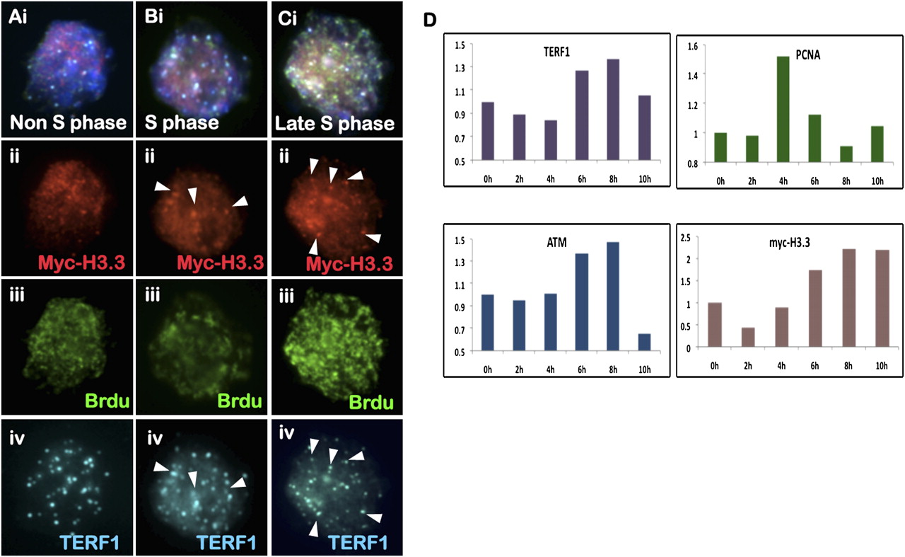
Cell cycle–dependent telomeric incorporation of MYC-H3.3. (A–C) Immunofluorescence analysis of ES129.1 cells expressing MYC-H3.3. MYC-H3.3 is induced by doxycycline for 12 h. No MYC-H3.3 (A, ii, red) was detected at the telomere in non-S-phase cells (negative BrdU-staining; A, iii, green). MYC-H3.3 (B, ii, red) was enriched at the telomeres (as shown by costaining with TERF1 [A–C, iv, blue]; arrowheads) in replicating S-phase cells (positive BrdU-staining; B, iii, green), with even more prominent telomeric signals detected by late S/G2 phase (C). (D) ChIP/real-time PCR analysis (n = 3; using primers specific to telomere) of synchronized (thymidine-blocked) ES129.1 cells using antibodies against TERF1, MYC tag, ATMS1981P, and PCNA. MYC-H3.3 expression was induced by doxycycline for 12 h. Data and calculations for the experiments are shown in Supplemental Table 2. An example of the ChIP/PCR experiment is shown in Supplemental Figure 11. PCNA association indicated an increase in replication activity at the telomeres following 2–6 h of release from thymidine-induced G1/S block. TERF1 association at the telomeres increased after 6 h post-G1/S-release. ATMS1981P association with the telomeres also increased during S phase. MYC-H3.3 incorporation at the telomeres occurred predominantly after 6 h of release at late S/G2 phase, in synchrony with the timing of telomere replication and processing.


