
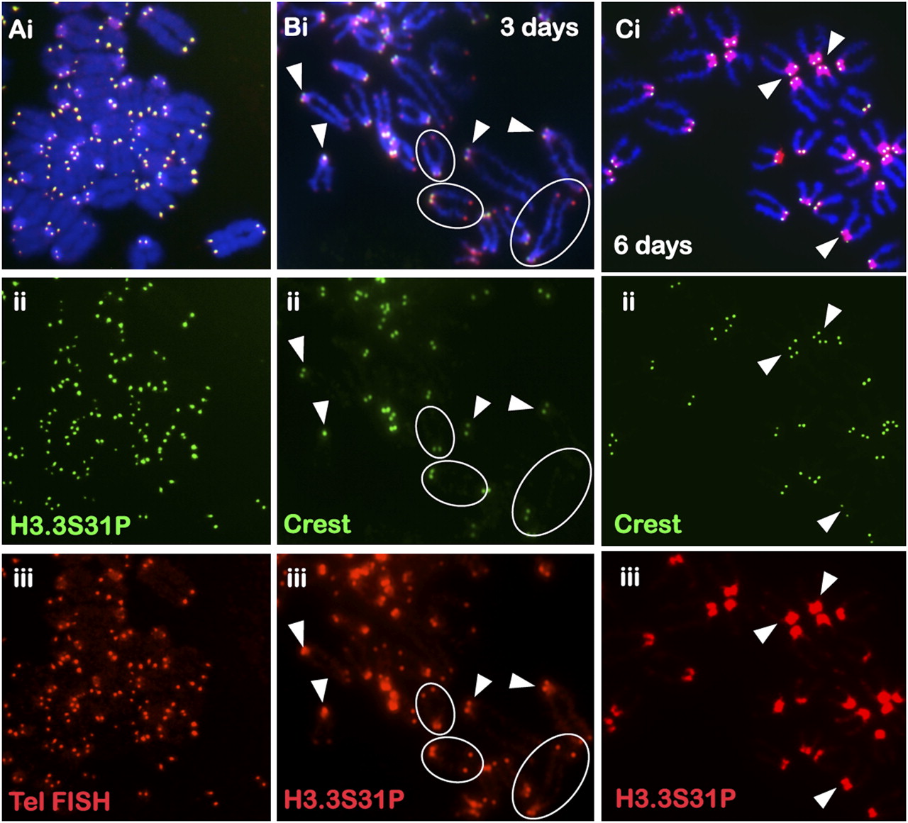
Figure 2.
Immunofluorescence analysis of H3.3S31P in mouse ES cells. (A) H3.3S31P (ii, green; Abcam) is enriched at the telomeres of all the chromosomes in ES129.1 cells, as confirmed by telomere FISH (iii, red). A second source of anti-H3.3S31P antiserum (Upstate; data not shown) gave a similar staining pattern. (B) Three days of differentiation resulted in decreased telomeric H3.3S31P signal (iii, red; some circled examples are shown) but enhanced staining at the pericentric DNA (arrowheads). (C) Six days of differentiation led to a near complete loss of telomeric H3.3S31P signals (iii, red) and a major increase in signals at the pericentric DNA. Centromeres were stained with CREST serum (B,C, ii, green).


