
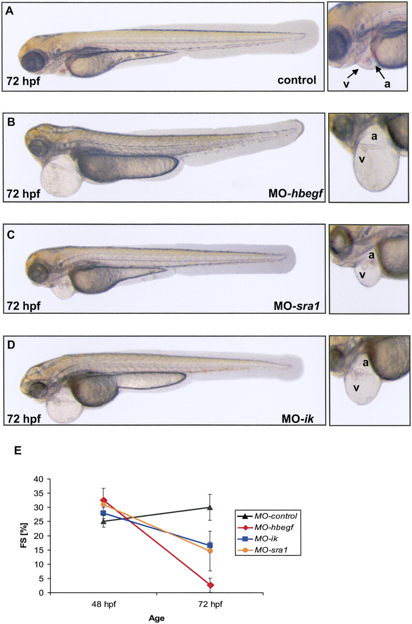
CM-cluster zebrafish knockdown phenotypes. Zebrafish embryos were injected with either control Morpholino (control) (A) or 2 ng of HBEGF Morpholino (MO-hbegf) (B), 4 ng of SRA1 Morpholino (MO-sra1) (C), or 2 ng of IK Morpholino (MO-ik) (D), and images were recorded at 72 hpf with a lateral view (head to the left, tail to the right). In contrast to the control-injected zebrafish embryos, hbegf, sra1, and ik morphants display severe pericardial edema. On the right, corresponding higher magnified views detailing cardiac chamber structure (a, atrium; v, ventricle) are shown for each morphant dysfunction. (E) Fractional shortening (FS) of the ventricular chamber was measured at different time points after injection (48 hpf and 72 hpf). Whereas in control-injected embryos, FS of the ventricle slightly increases from 48 hpf to 72 hpf, a dramatic decrease of FS in hbegf, sra1, and ik morphant ventricles can be observed by 72 hpf.


