
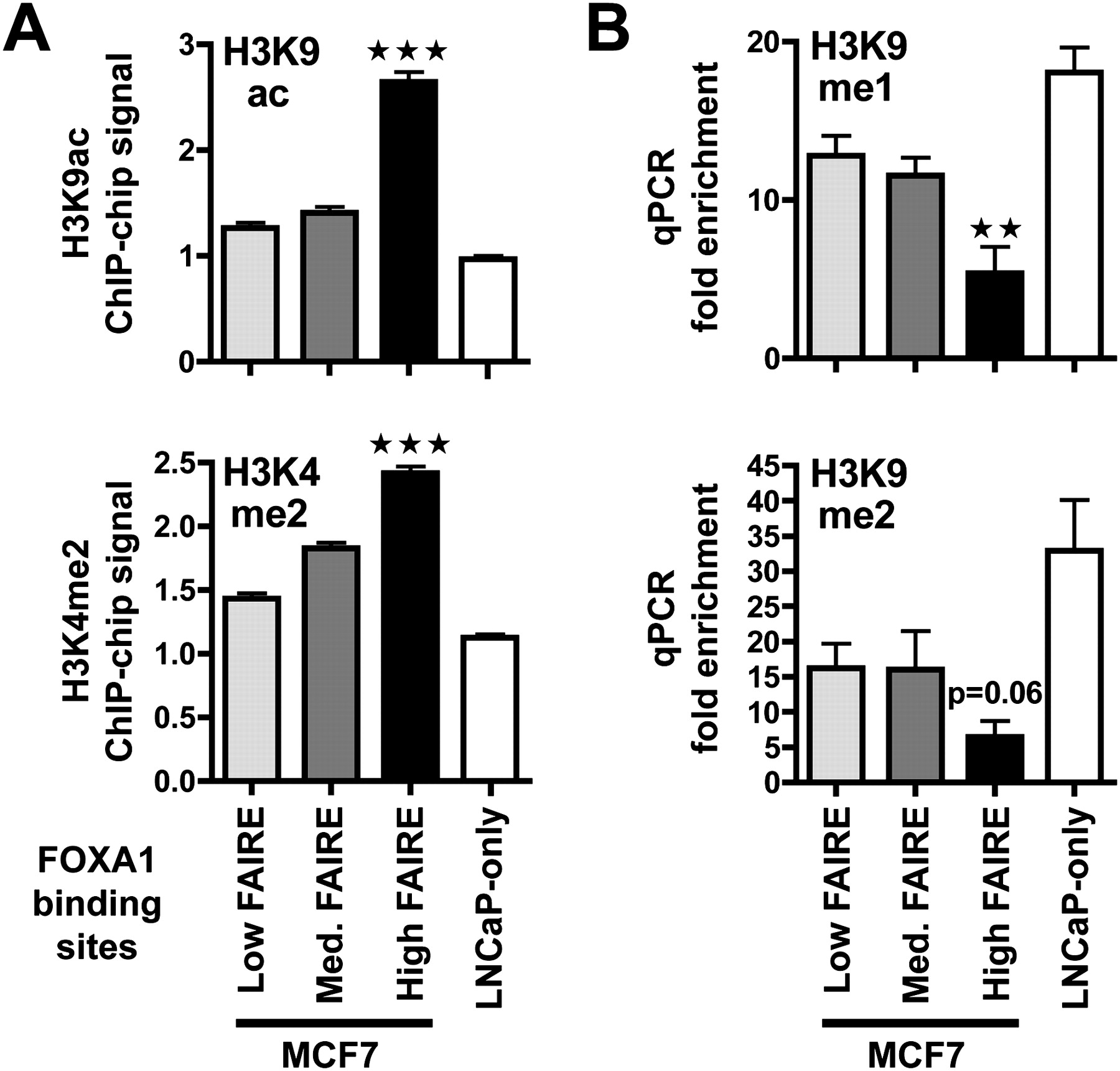
High FAIRE enrichment at FOXA1 binding sites correlate with a shift toward active histone marks. (A) H3K9ac and H3K4me2 ChIP-chip signals at FOXA1 binding sites from MCF7 or specific to LNCaP cells were analyzed. Data represent mean ± SEM of signals derived from MAT analysis of the ChIP-chip data. *** Indicates a statistically significant difference (P < 0.001) between FOXA1 sites from MCF7 with high FAIRE versus low FAIRE enrichment. (B) ChIP-qPCR experiments were performed to monitor H3K9me1 and me2 levels in MCF7 cells at the indicated categories of FOXA1 recruitment regions from MCF7 cells or specific to LNCaP cells (LNCaP only). Data show relative enrichments compared to negative control regions. Data are mean ± SEM from at least three independent experiments. Statistical significance of the difference between FOXA1 recruitment sites with high versus low FAIRE enrichments is shown (** indicates P < 0.01).


