
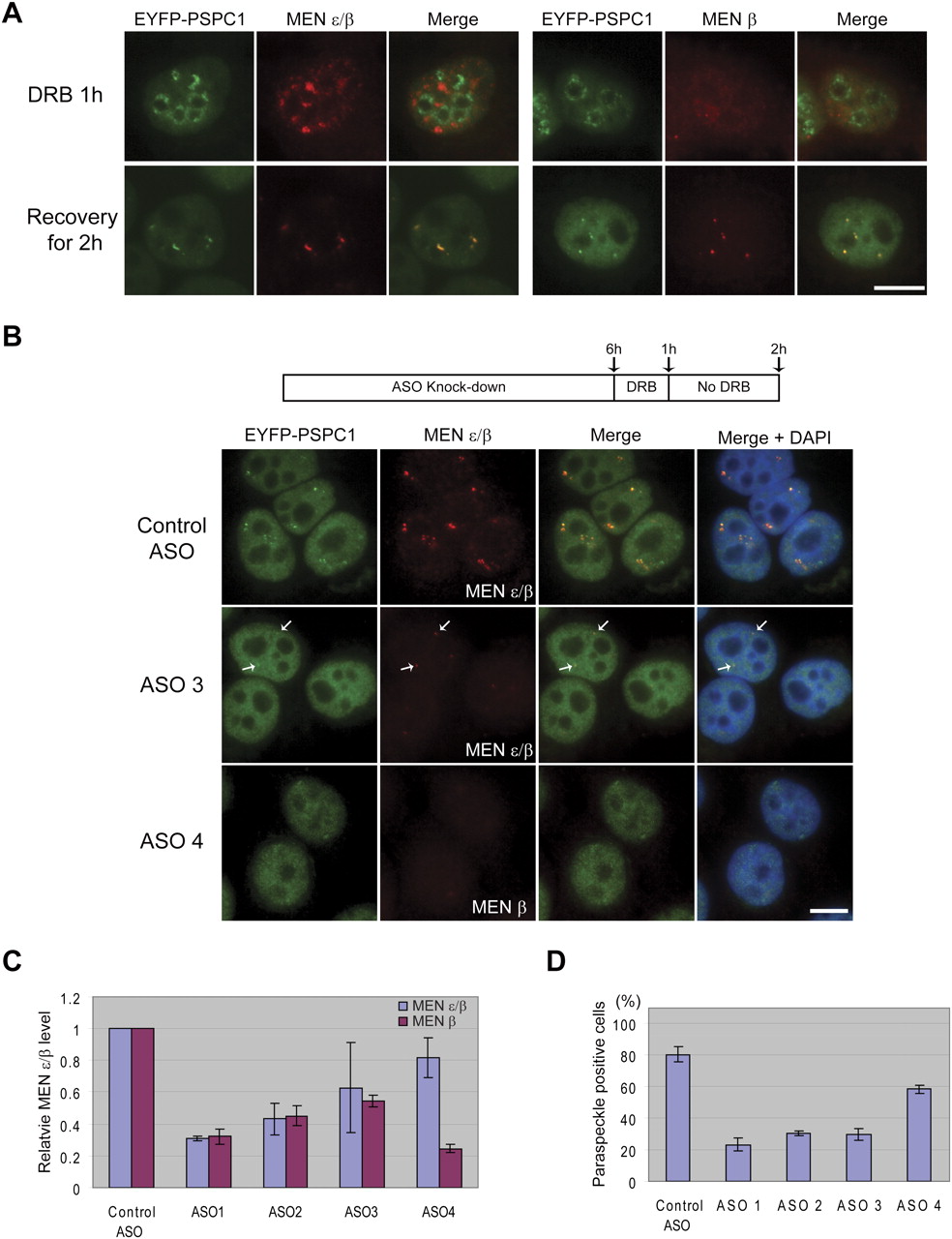
The reformation of paraspeckles after release from transcriptional inhibition is suppressed in MEN ε/β-depleted cells. (A) Upon DRB treatment for 1 h, EYFP-PSPC1 (also known as PSP1α) relocalized to the periphery of nucleoli. The MEN β transcript lost paraspeckle localization, while the MEN ε transcript relocalized to speckles. Upon removal of DRB and recovery for 2 h, paraspeckles reformed and colocalized with the MEN ε/β transcripts. (B) Cells were treated with ASOs to knock down MEN ε/β expression prior to DRB treatment and recovery. In cells treated with a control ASO, paraspeckles re-formed within 2 h of recovery. In contrast, paraspeckles did not re-form when MEN ε/β (ASO 3) or MEN β alone (ASO 4) was depleted. (Arrows) Residual paraspeckles in a cell where knockdown of the MEN ε/β transcripts was not complete. Scale bar, 10 μm. (C) Q-PCR was used to assess the ASO knockdown efficiency after 6 h of ASO treatment. A 40%–70% knockdown of MEN ε/β (ASO 1, 2, or 3) or ∼75% knockdown of MEN β (ASO 4) was achieved. Beta-actin was used as a normalization control. The data in the histogram are shown as mean and standard deviation values of three independent experiments. (D) The percentage of paraspeckle positive cells was reduced to 20%–30% by ASO 1, 2, or 3, while the control ASO did not influence the integrity of paraspeckles. Treatment with ASO 4 also resulted in a loss of paraspeckles, although to a lesser extent. The data in the histogram are shown as mean and standard deviation values of three independent experiments. Approximately 100 cells were counted per experiment.


