
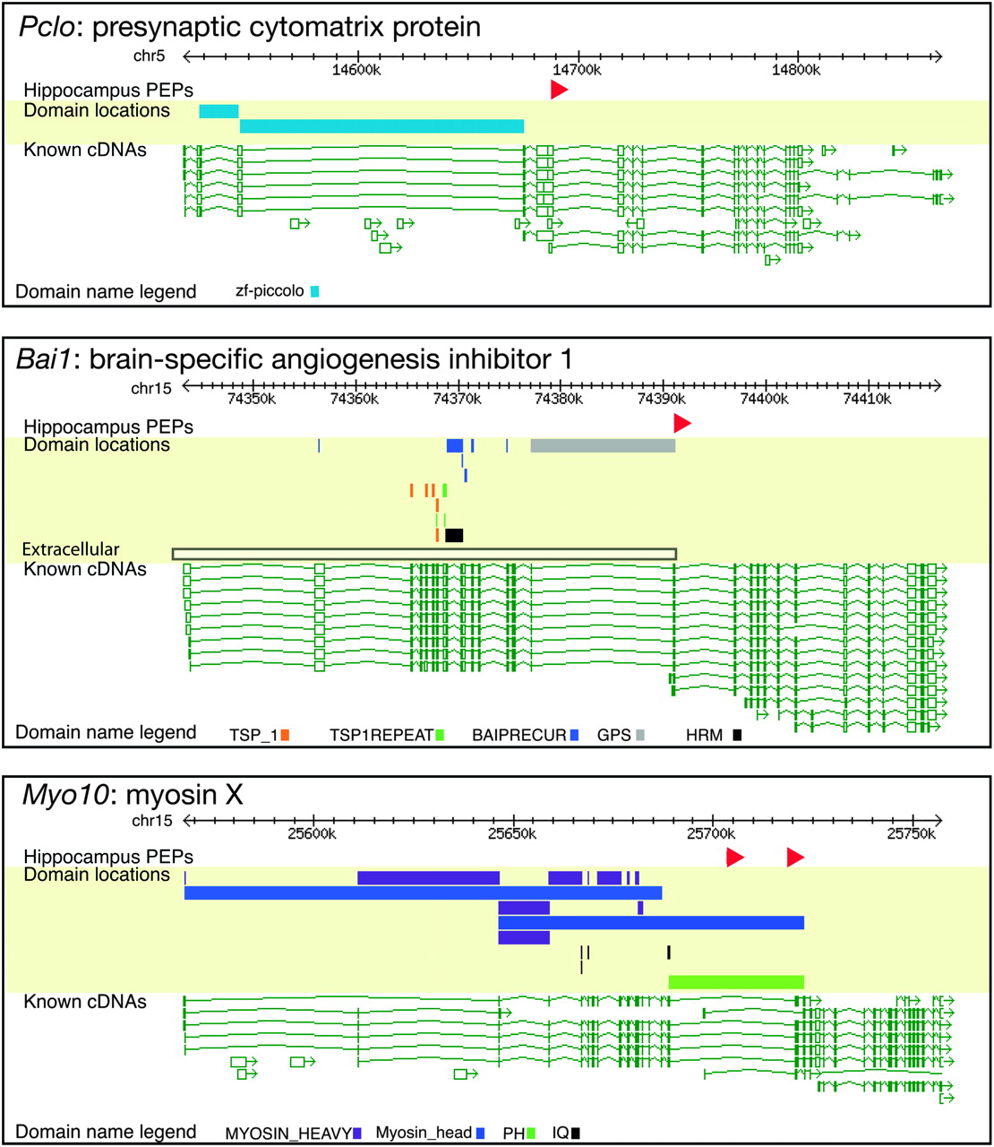
Examples of changes of domain content for genes by use of hippocampus PEPs. Hippocampus preferentially expressed promoter (PEP) locations are shown as red triangles. Locations of predicted protein domains are shown as colored blocks (note that domains spanning more than exons are extended over the intron(s). In all of these cases, at least one domain is upstream of the PEP, which means that this domain is not included in the isoform expressed in hippocampus. Known cDNA locations are shown below: transcription is right-to-left. BAI1 is a membrane protein whose N-terminal domain is extracellular, with a transmembrane region just downstream from the GPS domain (data not shown). The extracellular part can be cleaved off at the GPS domain, releasing a tumor-suppressing peptide; however, the hippocampus PEP is just downstream from the GPS domain, presumably giving a BAI1 variant that is attached to the membrane but without the extracellular domains, which lack the tumor-suppression capability (Kaur et al. 2005). Similarly, the PEPs in Myo10 confirm a previous study showing the neuronal expression of an isoform lacking the Myosin head domain (Sousa et al. 2006). In Pclo, the zf-piccolo domain cannot be included when using the hippocampus PEP.


